
The “Asynchronous Curtain Sign (ACS)” has been described as a finding in patients with pneumothorax (PTX). In my opinion, the ACS is the same as a previous clinical study in dogs that described the phenomenon as “Reverse A-lines” in a clinical PTX study out of Korea (Hwang et al. J Vet Sci 2028). The location for the screening for PTX is the “highest” region, least gravity dependent region, within the pleural cavity, referred to by the author as the TFAST® Chest Tube Site view, where “free air” in the pleural space would rise to in the presence of PTX.
The “Curtain Sign” of lung ultrasound represents the transition zone of pleural and abdominal cavities and is the covering and uncovering of the abdominal structures by a linear border, like a curtain, during phases of respiration in normalcy. The author always keeps cranial to the left on the screen and caudal on the right as in the Figure below. Because of the physics of ultrasound, air has a linear border again creating this covering and uncovering of abdominal structures in synchrony with phases of respiration.
This “Curtain Sign” of lung ultrasound also should be thought of as the transition zone of pleural cavity to the left off the screen and abdominal cavity to the right of the screen and is visible anywhere along this said interface of pleural cavity and abdominal cavities, i.e., Vet BLUE® Caudodorsal, Perihilar, and Middle Lung Regions. This “Curtain Sign” of lung ultrasound is analogous to the described phenomenon “Curtain Sign” of pleural effusion, where air filled lung covers and uncovers pleural effusion with phases of respiration.
To continue describing the “Curtain Sign” of lung ultrasound, the pleural side and abdominal side of this interface move very closely to being in synchrony during phases of respiration. With inspiration and expiration each side of the interface, this said transition zone, move together in synchrony. In PTX it has been described by Hwang et al., and Boysen et al., that the pleural side and abdominal side move asynchronously by moving toward one another and then away from one another during phases of respiration, inspiration and expiration, called “Reverse A-lines” by Hwang et al. and an “Asynchronous Curtain Sign” by Boysen et al., respectively.
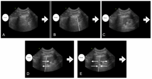
Figure by Lisciandro GR showing the “Curtain Sign” of lung ultrasound with the pleural air shadowing to the left, and abdomen being covered and uncovered to the right with the linear border unlabeled in A) during inspiration whereas the same image in B) has an overlay for the linear “curtain-like” border that in real-time would cover and uncover the abdominal structures in synchrony with phases of respiration; and when compared to C) expiratory (less coverage of the abdomen) the image in A) would be in inspiration (more coverage of the abdomen).
In C) the patient has expired (in expiration) and the liver in the near field could be mistaken for lung consolidation called the “Tissue Sign”, the rugal folds mid screen mistaken for lung consolidation called the “Shred Sign”, and far field the gastric contents with “Pseudo gastric B-lines” that can be mistaken for “wet lung” and types of alveolar-interstitial edema (Lisciandro’s mantra of “blood, water, or pus”). For these reasons it is important that the “Curtain Sign” of lung ultrasound be recognized and the probe moved cranially then from this transition zone of pleural and abdominal cavities to prevent such a misinterpretation.
In D) the pleural and abdominal sides move in synchrony with phases of respiration as indicated with the arrows, inspiration both move caudally, expiration both move cranially in synchrony.
In E) the pleural and abdominal sides are moving away from each other and then toward each other in asynchrony which is unexpected in normalcy and may be associated with PTX; however, other respiratory conditions that result in paradoxical abdominal breathing can create the asynchrony as well in the absence of PTX.
This is important for veterinary professionals using point-of-care ultrasound to be aware at triage since the finding of the “Asynchronous Curtain Sign” is not pathognomonic for PTX. Moreover, PTX is a “dry artifact” phenomenon, and thus A-lines would be present within the “Curtain Sign” of lung ultrasound without any “wet lung” artifacts, i.e., B-lines also called “lung rockets”, or any other findings of lung opposed to the thoracic wall during imaging, i.e., Vet BLUE® signs of consolidation called Shred, Tissue, Nodule, and Wedge Signs.
A Clinical Algorithm has been created by Lisciandro GR.
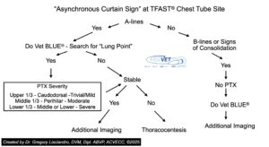
Here is a short video showing examples of “synchronous” and “asynchronous curtain signs” of lung ultrasound with case examples. THIS VIDEO WILL PLAY but has a SHORT SEVERAL SECOND DELAY and lasts about 90 seconds. Enjoy the didactic video short. THERE is NO SOUND. As you watch, apply the clinical algorithm above…
Here is a PDF of then Poster Abstract presented at IVECCS 2025

grl/GLR 9-8-2026