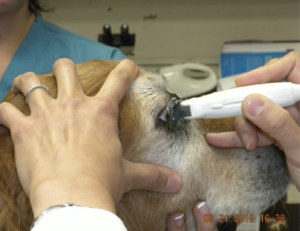
Courtesy of Dr. Jane Cho, DVM, DACVO, Point-of-care Ultrasound for the Small Animal Practitioner, 2nd Edition, Wiley-Blackwell, © 2021.
Imaging the Eye – Ocular Ultrasound – It’s So Easy Any Caveman or Cavewoman Could Do It! – Lisciandro GR
Imaging the eye is the best kept secret of point-of-care ultrasound. Why? Because the eye is fluid filled, just like the urinary bladder and the gallbladder for examples, and of note as well, ultrasound’s strength is imaging through fluid! The major critical aspect of imaging the eye is using a safe coupling medium that will not injure the cornea. The major contraindication is not to image an eye with a ruptured globe. Watch our FASTVet Awesome Webinar – It’s linked below…
I learned how to image the eye by reading Dr. Jane Cho’s chapter in the 1st edition of our textbook that is updated in our 2nd edition, Point-of-care Ultrasound for the Small Animal Practitioner, Wiley-Blackwell, © 2021. Dr. Jane Cho, DVM, DACVO is a veterinary board-certified veterinary ophthalmologist that practices in Westchester County near New York City. I have used the linear probe as well as the microconvex probe that she uses in her book chapter. Click here.
Briefly, the eye is first anesthetized and a topical ocular anesthesia protocol is outlined in Dr. Cho’s chapter. If in a pinch, you can always use sterile K-Y Jelly as your coupling medium. I always open a new unopened tube or packet so I know that I will not transfer any bacteria or foreign material onto the eye. K-Y Jelly is safe but its disadvantage is that K-Y Jelly doesn’t have the “glob” of holding power that say Aquasonic commercially available ultrasound gel would have. However, always check with the manufacturer’s recommendations to make sure that whatever coupling medium used is safe for the eye.
Watch our Imaging the Eye – Ocular Ultrasound Webinar. Take great pride in learning this skill because very few non-opthalmologists and non-imaging specialists (meaning veterinarians that are not radiologists) are providing this service for their patients. When I was practicing in the ER, I would text my ocular ultrasound images to our local veterinary ophthalmologist (and he really liked this process) to get an idea if the eye required urgent care or could wait until the morning.
For example, “Dr. Eye” as I’ll call him, would be out for a dinner with his wife and friends, and would be able to look at my ocular imaging information and decide on how soon the patient needed to be seen. This was also important for the client, paying an emergency fee versus scheduling an appointment for the next day. I think you get my point.
This is our Awesome FASTVet webinar:
FASTVet Monthly Webinar October 2020: Ultrasounding the Eye – It’s So Easy
This is also very nicely done for human patients and an article with images that can be translated to our veterinary patients covering ocular ultrasound in people:
Send your comments to Dr. Gregory Lisciandro, DVM, DABVP, DACVECC to FastSavesLives@gmail.com or LearnGlobalFast@gmail.com. We hope you find this Blog and the associated links helpful and learn this skill in addition to Global FAST®!
gl/GL 11-1-2024