Ultrasound Assessment of the Gallbladder Wall: An Important Marker in the Acute Setting
Stephanie Lisciandro, DVM, DACVIM (SAIM)
Ultrasound assessment of patients is extremely important in the acute veterinary setting. Point-of-care ultrasound (Global FAST®) should be used in addition to thorough physical exam and laboratory evaluations to offer the timeliest diagnosis and therapeutic intervention in critical patients. Ultrasound assessment of the gallbladder has proven to be an important tool. The finding of gallbladder wall edema (GBWE) in acutely ill patients can be a marker for anaphylaxis, right-sided heart failure, pericardial effusion, or acute cholecystitis. Other documented causes include hypoproteinemia, immune-mediated anemia, sepsis, pancreatitis, and post-transfusion (likely immune-mediated; possibly volume overload).
In this discussion, we will focus on gallbladder wall edema in patients with cardiac disease. This was reviewed in a recent article in the Journal of Veterinary Internal Medicine entitled, “Thirteen dogs and a cat with ultrasonographically detected gallbladder wall edema associated with cardiac disease” (JVIM 2021; Lisciandro GR, et al). Knowing this significant ultrasound finding is especially important in the emergent setting since patients with anaphylaxis and cardiac disease may have similar presentations including acute weakness and collapse.
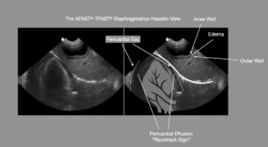
Gallbladder wall edema (GWBE). Pericardial effusion can be seen as an anechoic rim around the heart called the “Racetrack Sign” at the AFAST®-TFAST® Diaphragmatico-Hepatic (DH) View on Global FAST® exam.
It in this study, GBWE is described as an ultrasonographically striated gallbladder wall with mural thickening ranging from 3-5 mm – a white-black-white or white-gray-white monographically appearing striation. This finding is also commonly known as the “gallbladder halo sign.”
Of the dogs in our published peer-reviewed case series, 11 of the 13 dogs had pericardial effusion, one had dilated cardiomyopathy and one had right ventricular myocardial failure. In addition, 9 of the 13 dogs had ascites. One cat was also included in study and this patient had right sided heart failure associated with a ventricular septal defect; and the cat also had ascites.
In patients with acute weakness and/or collapse, ultrasonographic assessment of the gallbladder is very important because cardiac causes of GBWE and canine anaphylaxis can present similarly. Patients with ascites are often presented for abdominal ultrasound and as a result thoracic pathology is missed since these animals often do not have heart murmurs or other findings indicating thoracic ultrasound (lung, cardiac, or both). It is important to remember to look past the diaphragm with enough depth to see into the thorax at the AFAST®-TFAST® Diaphragmatico-Hepatic View when evaluating the liver to look for pericardial effusion. It is also important to assess these animals for distention of the caudal vena cava and hepatic veins – see CVC blog.
In dogs with a “gallbladder halo sign” secondary to anaphylaxis (unique to dogs having their shock organ liver and gastrointestinal tract) referred to as an “anaphylactic gallbladder”, the caudal vena cava is often small or collapsed (“flat”) due to decreased venous return from severe life-threatening hypovolemic, distributive shock. A distended vena cava (“FAT”) with lack of “bounce” (due to back flow of blood from a failing right heart or obstruction of flow from pericardial effusion), supports the presence of cardiogenic GBWE referred to as a “cardiac gallbladder.” The point-of-care protocol for Global FAST® (AFAST®, TFAST®, and Vet BLUE® combined – the video) automatically incorporates a standardized assessment of all of these findings to allow a more accurate diagnosis of pericardial effusion or right sided heart failure versus other causes of GBWE.
Ultrasound assessment is a valuable tool for the assessment especially in the emergent setting, but I would advise ultrasound assessment in any sick animal as an extension of the physical exam. Please sign up for our emails or join the FASTVet.com for additional resources regarding using ultrasound every day in your practice.
References:
- Lisciandro GR, Gambino JM, Lisciandro SC. Thirteen dogs and a cat with ultrasonographically detected gallbladder wall edema associated with cardiac disease. J Vet Intern Med 2021;35: 1342–1346. https://doi.org/10.1111/jvim.16117.
- Chou Y, Ward JL, Baron LZ, Murphy SD, Topf MA, Lisciandro GR, et al. Focused ultrasound of the caudal vena cava in dogs with cavitary effusions or congestive heart failure: a prospective observational study. PLoS One 2021;16(5):e0252544. doi:10.1371/journal.pone.0252544.
- Lisciandro GR. Abdominal and thoracic focused assessment with sonography for trauma, triage and monitoring in small animals. J Vet Emerg Crit Care 2011;21(2):104-122.
- Lisciandro GR. The use of the diaphragmatico-hepatic (DH) views of the abdominal and thoracic focused assessment with sonography for triage (AFAST/TFAST) examinations for the detection of pericardial effusion in 24 dogs (2011-2012). J Vet Emerg Crit Care 2016; 26(1):125-31.
- Nelson NC, Drost WT, Lerche P, et al. Noninvasive estimation of central venous pressure in anesthetized dogs by measurement of hepatic venous blood flow velocity and abdominal venous diameter. Vet Radiol Ultrasound 2010;51(3):313-323.