To learn Vet BLUE®, take our 7-hour Online RACE-approved Course. If you can image the urinary bladder, you can learn Vet BLUE® and immediately improve patient care! Click here for more information and to sign up!
I’ve had the privilege of listening to lectures presented at the TRISAT Critical Care Webcast Series over the past decade that are webcasted throughout the state of Texas including Level 1 trauma centers (and I have been an invited lecturer 3 times to this group at one time lecturing more to them than at veterinary conferences).
I have heard through this group as well as other internationally renowned lung ultrasound sonographers including Dr. Enrico Storti “that lung ultrasound is almost as good as computed tomography (CT) for wet lung conditions.” In fact, Dr. Storti made this statement at the World Congress of Veterinary Anesthesiology (WCVA) in Italy several years ago (2018) where I too was a speaker.

Picture of Dr. Storti, MD (taller) and me Dr. Lisciandro, DVM (shorter) as 2 Italian lung sonographers!
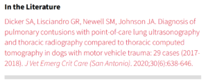
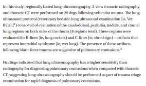
The only study that I am aware of in veterinary medicine that compares “Wet Lung” with the 3 major imaging modalities of thoracic radiography (TXR) to lung ultrasonography (LUS) to the reference standard computed tomography (CT) is by Dicker, Lisciandro, Newell, and Johnson published in 2020. Pulmonary contusions are “Wet Lung” conditions with blood (wet) being cuffed by aerated alveoli. “Wet lung” is characterized by the B-line or ultrasound lung rocket artifacts. “Wet lung” artifacts are nonspecific, meaning that “Wet Lung” could be water, blood, or pus cuffed by aerated alveoli at the lung surface. Because they are nonspecific, “Wet Lung” is often referred to as alveolar – interstitial syndrome (AIS). There are less common causes of “Wet Lung”; however 95% of the time in small animals (author opinion), “Wet Lung” (B-lines, lung rockets are synonymous) is due to some form of alveolar – interstitial edema (i.e., water, blood, pus).
In trauma, “Wet Lung” represents lung contusions until proven otherwise. In the Dicker et al. study, these 3 imaging modalities were acquired within 30-minutes in the order of TXR 3-view to Vet BLUE® to the reference standard of CT. Click here for the link to the study’s abstract in pubmed.
The findings support the statement by Dr. Enrico Start, MD, lung ultrasound expert, that “Lung ultrasound is almost as good as CT for “Wet Lung” conditions.” Dicker et al. found that with CT as the reference standard and 100%, Vet BLUE® performed at 92% and TXR at 67%.
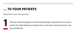
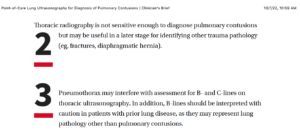
Let’s look at the Clinician’s Brief article from just a few weeks ago that comments on our study. The review was written by Dr. Britt Thevelein, DVM, DACVECC. Click here for the link. See below for some of its excerpts:
This FASTVet Blog was written by Dr. Greg Lisciandro, DVM, DABVP, DACVECC. You may contact Dr. Lisciandro by email at LearnGlobalFAST@gmail.com or by cell at 210-260-5576. Excerpts from Clinician’s Brief were used with their permission.
To learn Vet BLUE®, take our 7-hour Online RACE-approved Course. If you can image the urinary bladder, you can learn Vet BLUE® and immediately improve patient care using our 2-minute exam!
Click here for more information and to sign up for our Online Vet BLUE® Course!
For a full list of our publications including Vet BLUE®, go to the lead page of FASTVet, scroll down, and click on “More Publications” or use this LINK.