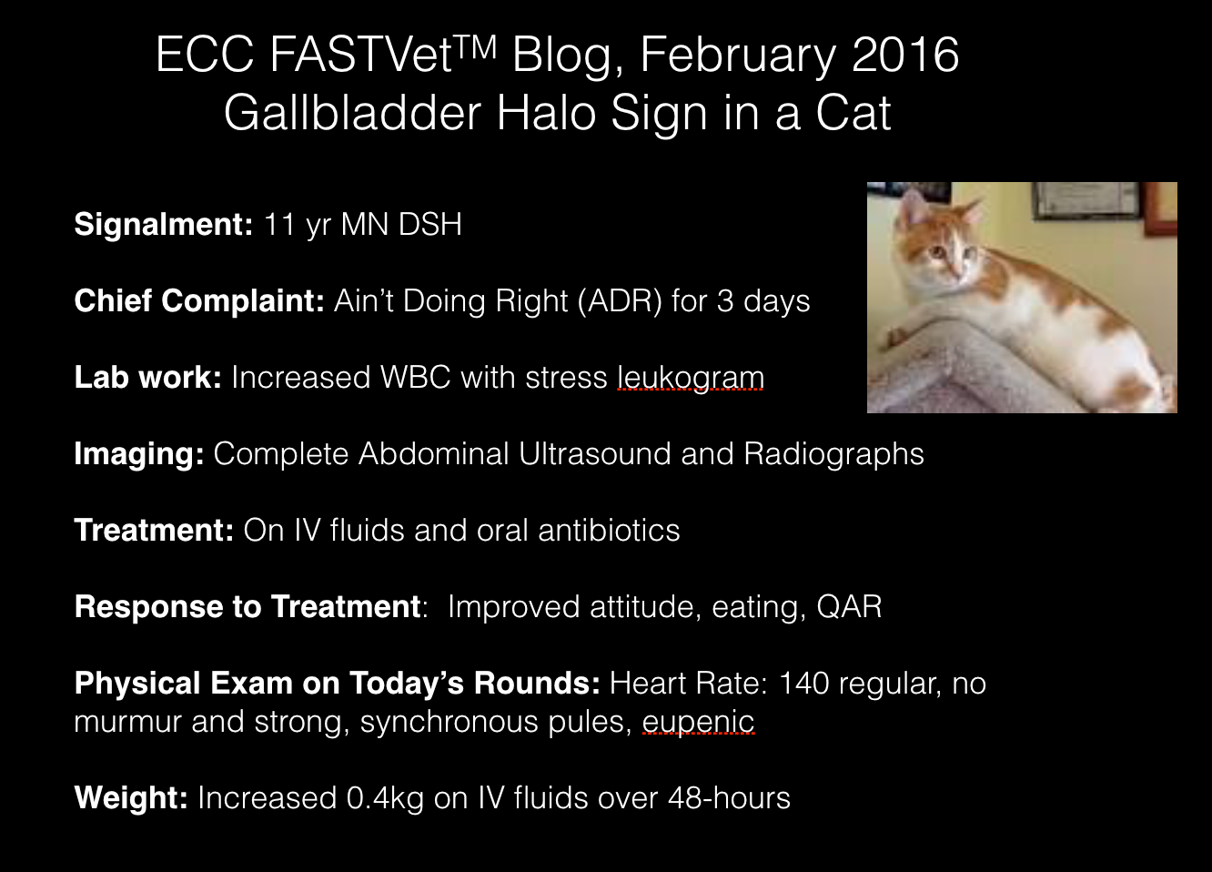
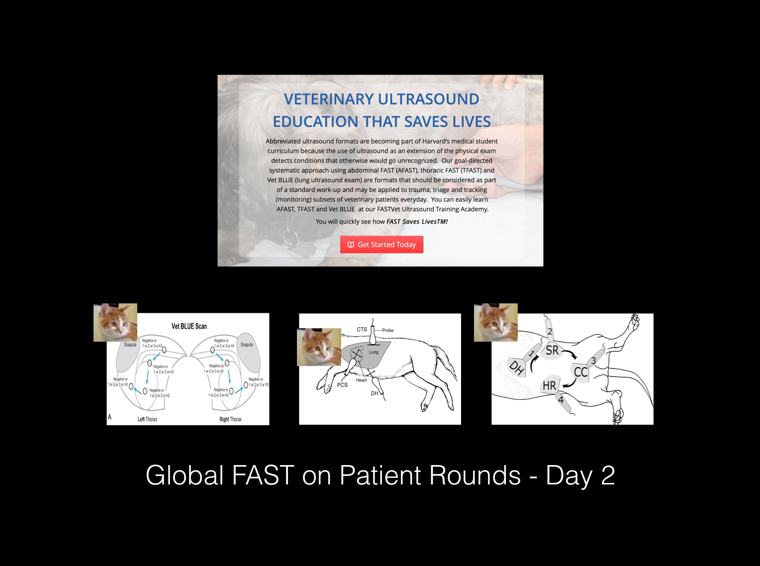
This is a great case from 2016 but we think you will find it very interesting! This case illustrates an important ultrasound finding, this time in the feline species (for all the FASTVet.com cat lovers!) We describe Gallbladder Wall Edema (the “Gallbladder Halo Sign”) related to heart failure in a cat!
The case also demonstrates how Global FAST® gives you information that you might miss with traditional ultrasound formats such as Complete Abdominal Ultrasound and Complete Echocardiography.
Click on Image to Make Larger
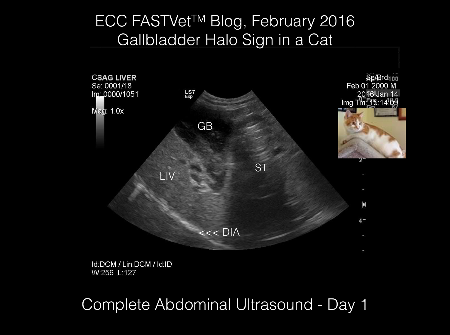
Eddie is an 11yr old, MN Domestic Shorthair (DSH) with a 2-day history of lethargy and hiding. Complete Abdominal Ultrasound performed by the owner of the practice reveals the following:
- An unremarkable liver (LIV: liver; GB: gallbladder; DIA: diaphragm; ST: stomach) -next 4 images
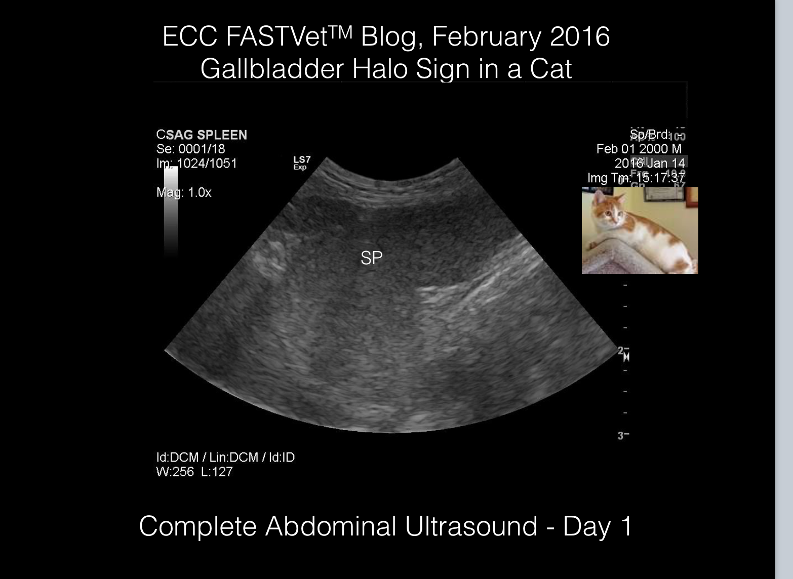
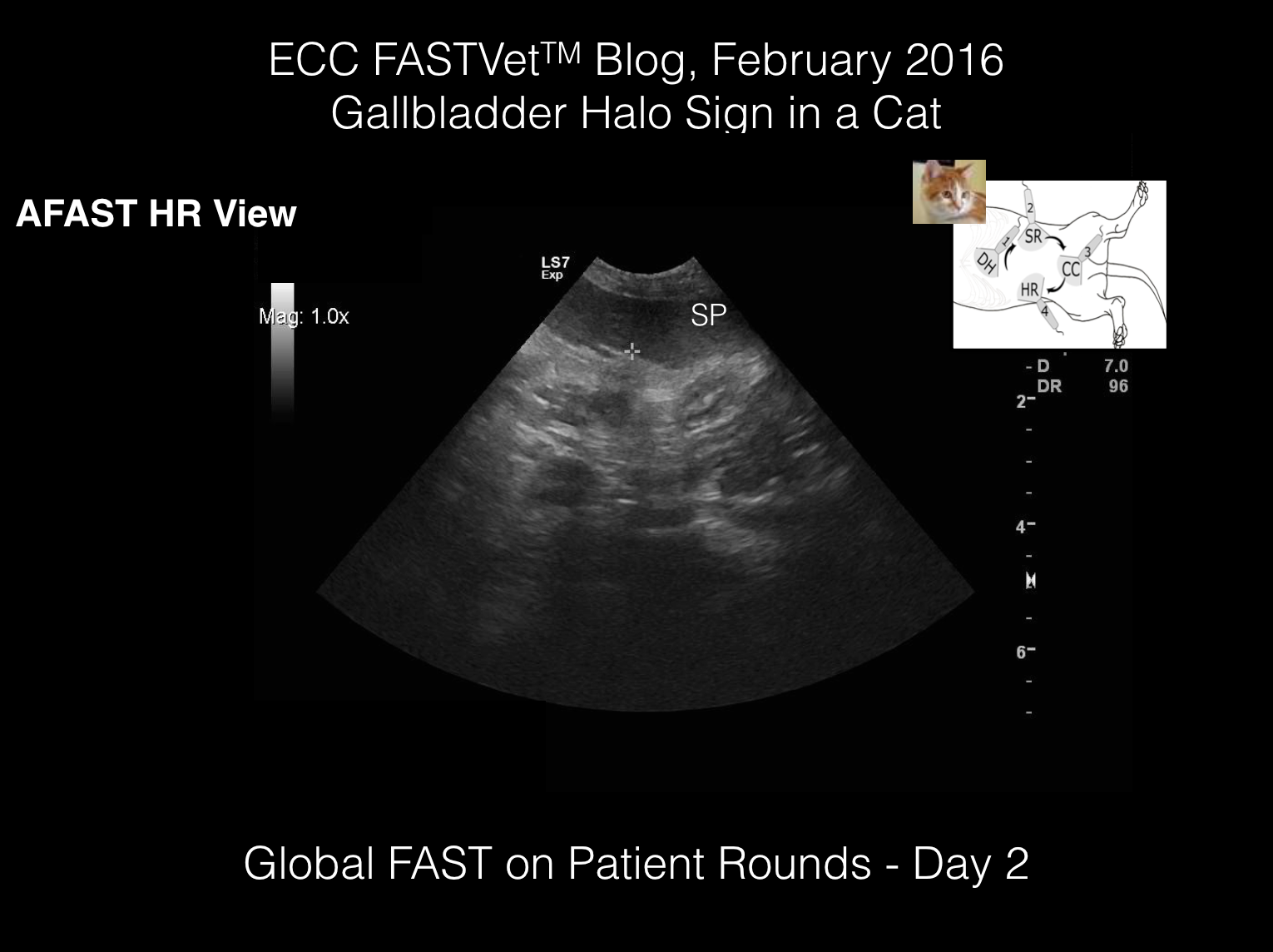
- An unremarkable spleen (SP: spleen), although it appears to be enlarged/thickened (cat normal < 10 mm)
- A urinary bladder (UB: urinary bladder) that appears to have echogenic sediment
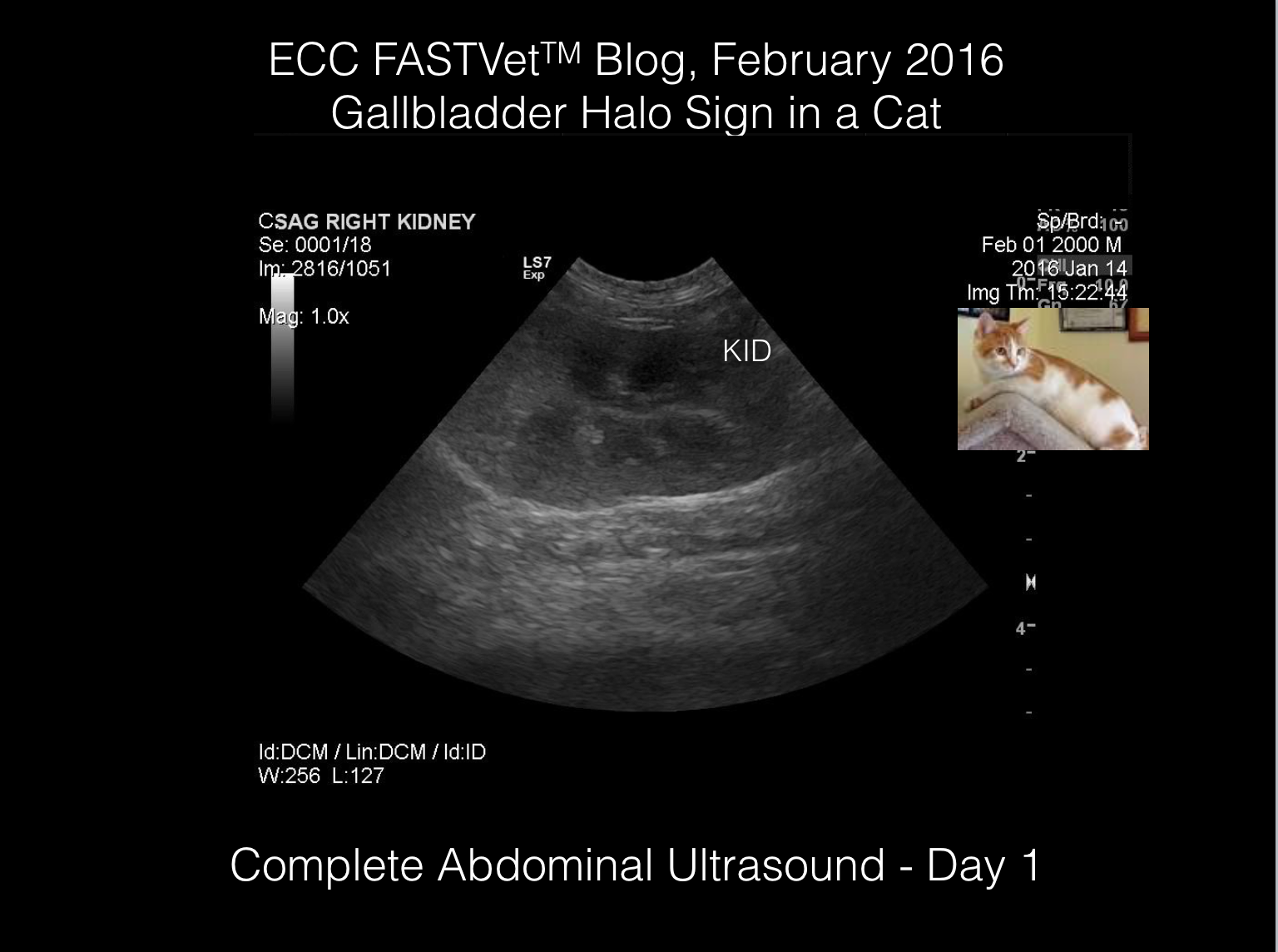
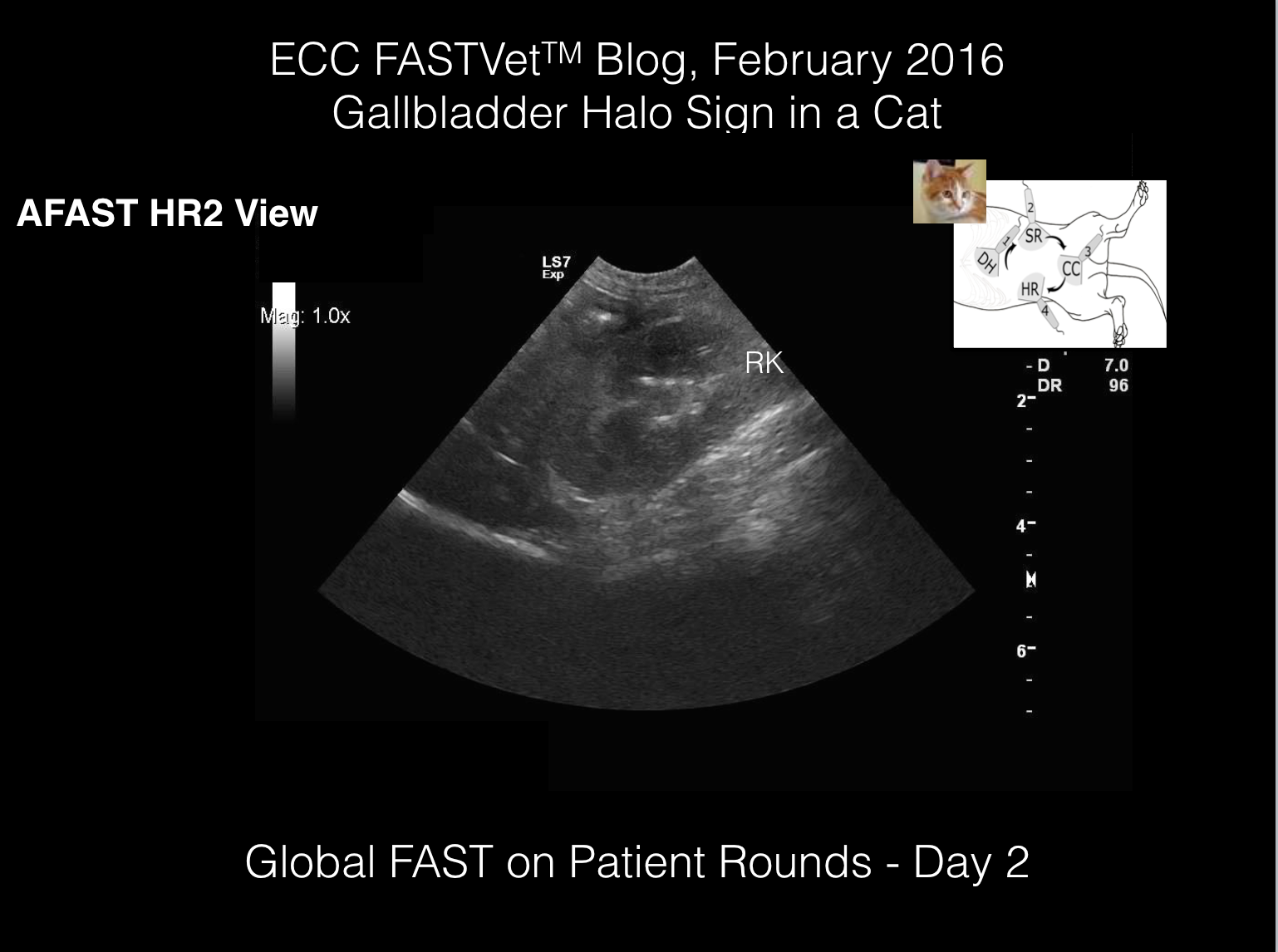
- An unremarkable left and right kidney (only 1 shown)
Click on Image to Make Larger

Click on Image to Make Larger

Click on Image to Make Larger

Click on Image to Make Larger
- An unremarkable liver (LIV: liver; GB: gallbladder; DIA: diaphragm; ST: stomach)
- An unremarkable spleen (SP: spleen), although it appears to be enlarged/thickened (cat normal < 10 mm) – next 2 images
- A urinary bladder (UB: urinary bladder) that appears to have echogenic sediment
- An unremarkable left and right kidney (only 1 shown)
Click on Image to Make Larger

Click on Image to Make Larger
- An unremarkable liver (LIV: liver; GB: gallbladder; DIA: diaphragm; ST: stomach)
- An unremarkable spleen (SP: spleen), although it appears to be thickened (cat normal < 10 mm)
- A urinary bladder (UB: urinary bladder) that appears to have echogenic sediment – next 2 images
- An unremarkable left and right kidney (only 1 shown)

Click on Image to Make Larger

- An unremarkable liver (LIV: liver; GB: gallbladder; DIA: diaphragm; ST: stomach)
- An unremarkable spleen (SP: spleen), although it appears to be thickened (cat normal < 10 mm)
- A urinary bladder (UB: urinary bladder) that appears to have echogenic sediment
- An unremarkable left and right kidney (only 1 shown) – next image
Click on Image to Make Larger
So Assessment is summarized as a cat with an elevated WBC, active urinary sediment (with importantly unremarkable kidneys), with otherwise unremarkable abdominal imaging. The working diagnosis is a urinary tract infection (UTI). Eddie is treated with intravenous fluids at x3 maintenance rate, and a once a day fluoroquinolone antibiotic.
Click on Image to Make Larger
The Global FAST® (combination of AFAST®, TFAST® and Vet BLUE®) format we have developed, is used as a first line imaging modality (trauma, non-trauma) and for tracking (monitoring cases). Global FAST® is used during delay patient rounds because it assesses patient volume status and detects complications otherwise missed by physical examination, lab work, radiography AND Complete Abdominal Ultrasound. Global FAST® is a different mindset than traditional Complete Abdominal Ultrasound and Complete Echocardiography.
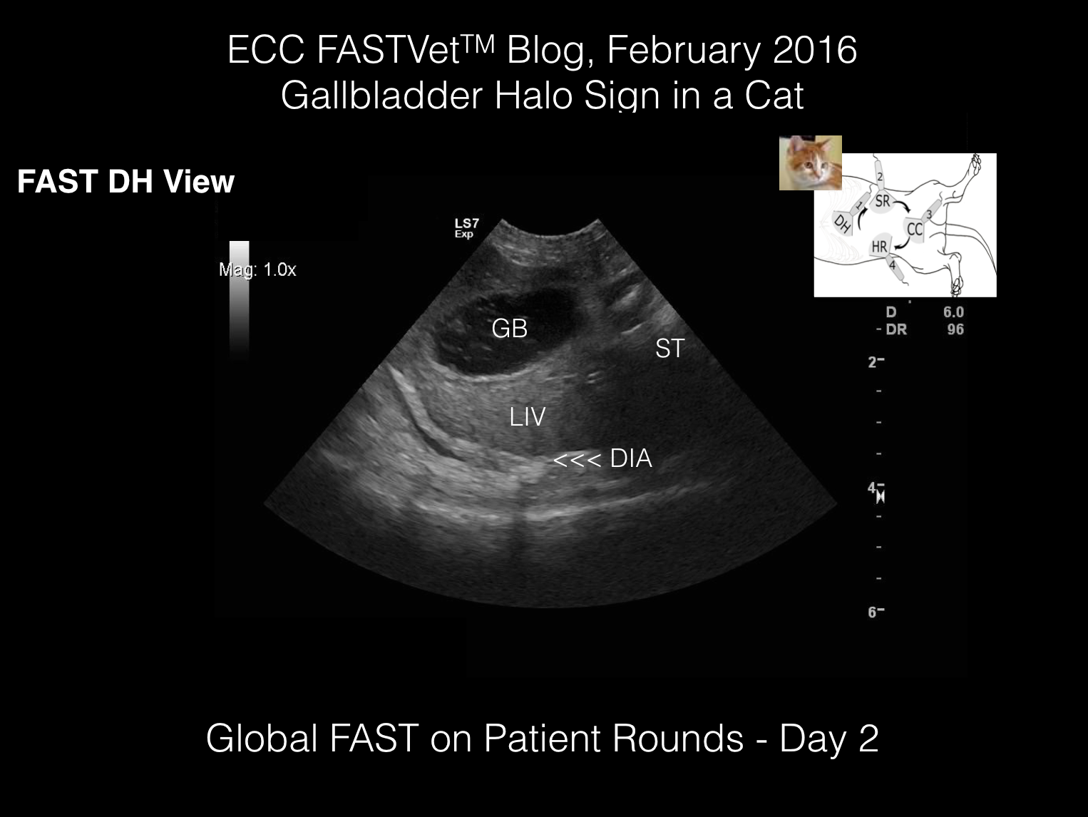
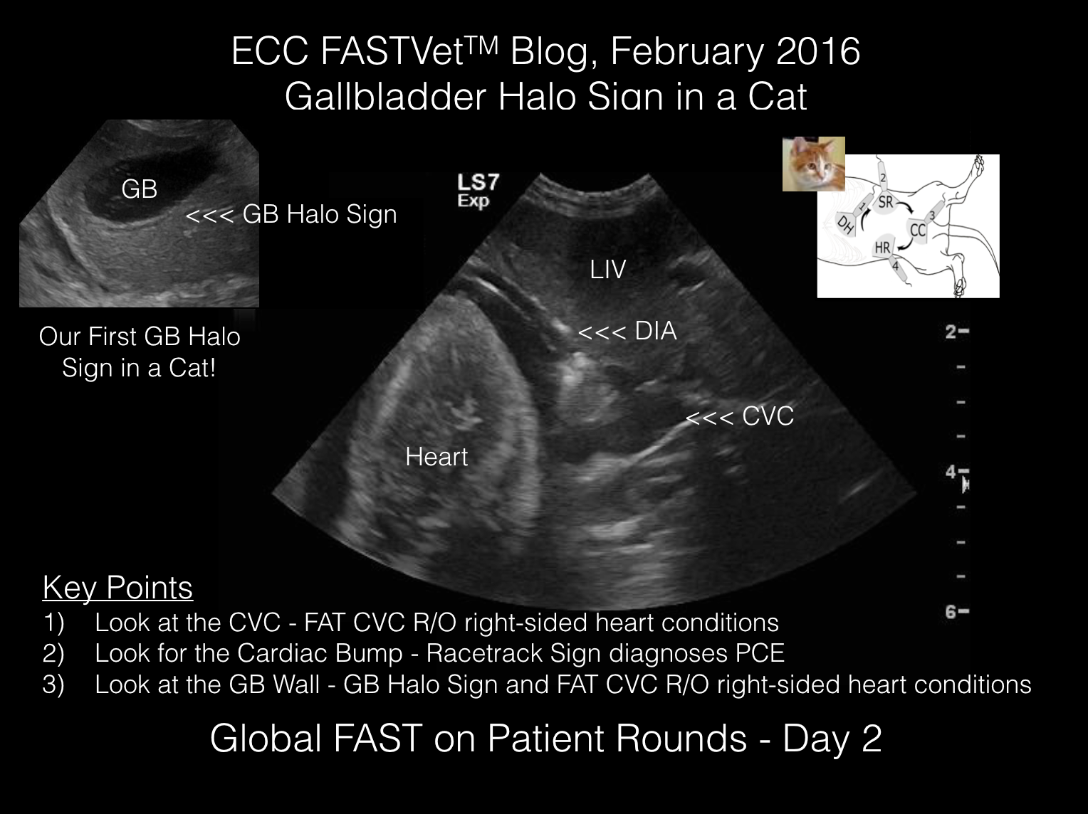
What we find on Day 2 using Global FAST immediately after our physical examination:
- Gallbladder Wall Edema (referred to as the “Gallbladder Halo Sign”)
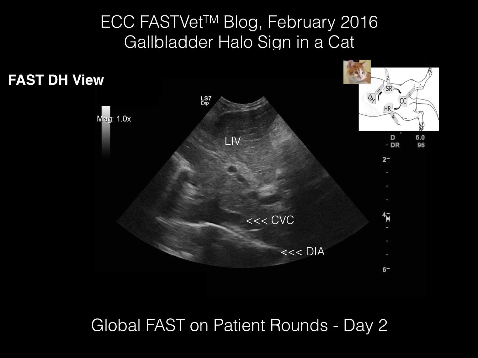
- Caudal Vena Cava (CVC) and hepatic venous distension
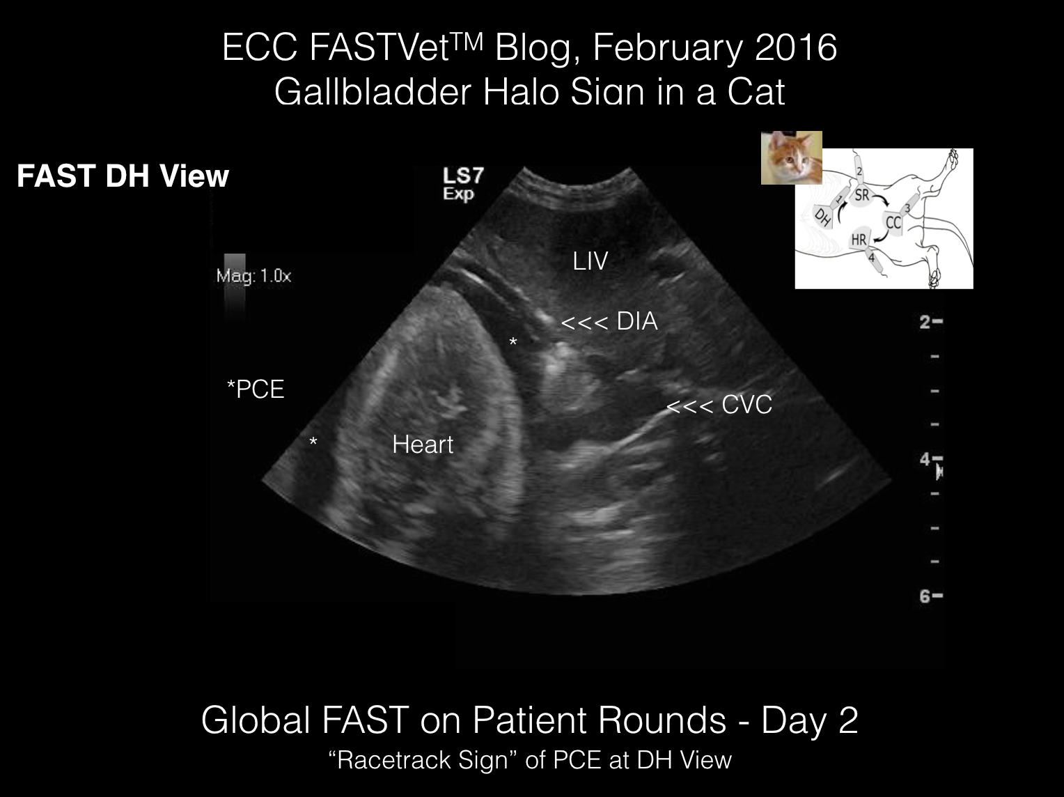
- Pericardial Effusion via the FAST DH View
Our conclusion here, and based on increased body weight, is that Eddie is not tolerating the IV fluids and the right heart is failing (overloaded). Clinically Eddie feels better, and physical examination, lab work, and radiographic imaging are insensitive to detect the complication (at least in its beginnings).
- Gallbladder Wall Edema (referred to as the “Gallbladder Halo Sign”)
- Caudal Vena Cava (CVC) and hepatic venous distension
- Pericardial Effusion via the FAST DH View
- Gallbladder Wall Edema (referred to as the “Gallbladder Halo Sign”)
- Caudal Vena Cava (CVC) and hepatic venous distension
- Pericardial Effusion via the FAST DH View
- Gallbladder Wall Edema (referred to as the “Gallbladder Halo Sign”)
- Caudal Vena Cava (CVC) and hepatic venous distension
- Pericardial Effusion via the FAST DH View
Click on Image to Make Larger
The remainder of the AFAST® exam is unremarkable at the SR, CC, HR and HR2 Views.

Click on Image to Make Larger

Click on Image to Make Larger
Click on Image to Make Larger
Click on Image to Make Larger
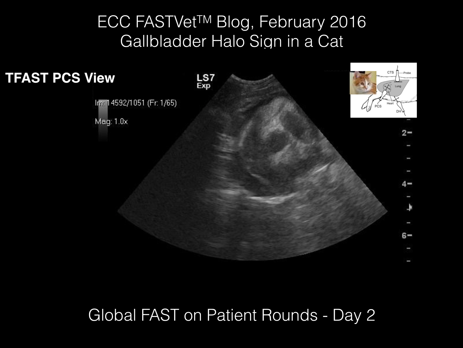
The TFAST® PCS View can be confusing for the diagnosis of Pericardial Effusion unless the sonographer images towards the muscular apex of the heart where no heart chambers can be confused with PCE (or pleural effusion).
The next view is too high toward to the base of the heart and a dangerous manner in which to diagnose PCE.
Click on Image to Make Larger
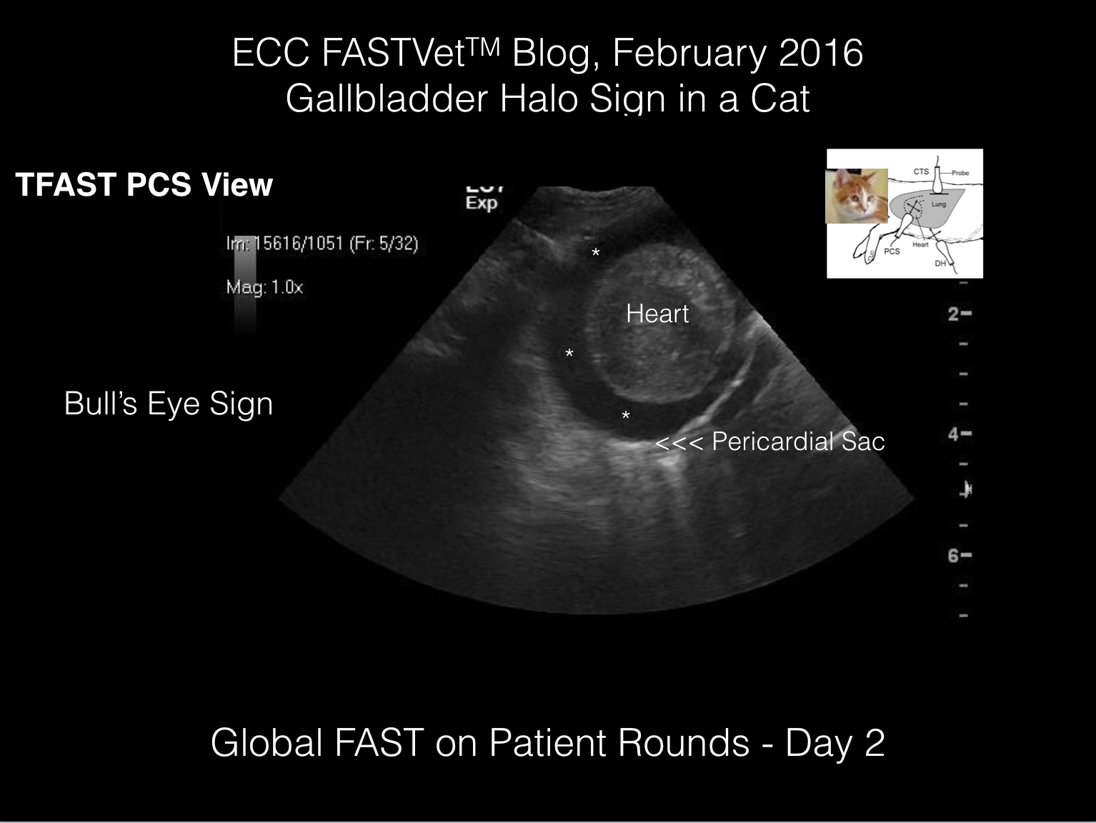
The view below shows how imaging toward the muscular apex of the heart, similar to the FAST DH View’s Racetrack Sign, reduces the mistaking of a heart chamber for an effusion. At the TFAST® PCS View, the Bull’s Eye Sign is viewed when PCE is present.
Click on Image to Make Larger
By incorporating Vet BLUE®, left-sided heart function can be rapidly assessed by the presence or absence of B-lines or the Wet vs. Dry Lung Principle. Eddie is DRY ALL Vet BLUE Fields so diuretics are unnecessary (or should be used very judiciously). By completing Global FAST, the Assessment is that Eddie is sub-clinically experiencing right-sided heart failure/volume overload and needs to have his IV fluids discontinued.
Complete Echocardiography is indicated in an ideal world; however, knowing that congestive heart failure (here only right-sided is occurring [lung is DRY]) is the most common cause of pericardial effusion in cats, monitoring with daily Global FAST® expecting resolution of PCE may all that is needed in Eddie’s short-term course. Eddie has favorably responded to initial therapy and no each decisions should be made (don’t doom Eddie).

Click on Image to Make Larger
KEY POINTS:
- Complete Abdominal Ultrasound tends to miss intra-thoracic conditions by not looking into the pleural and pericardial space
- Complete Abdominal Ultrasound tends to miss right-sided volume overload unless the sonographer is trained to pay attention to the caudal vena cava and hepatic veins for signs of hepatic venous congestion (FAT CVC, distended hepatic veins [Tree Trunk Sign])
- Gallbladder Halo Sign although rare in cats, does serve as a marker for right-sided heart conditions as it reliably does in dogs and humans (although be sure to know other rule outs for Gallbladder Halo Sign)
Click on Image to Make Larger
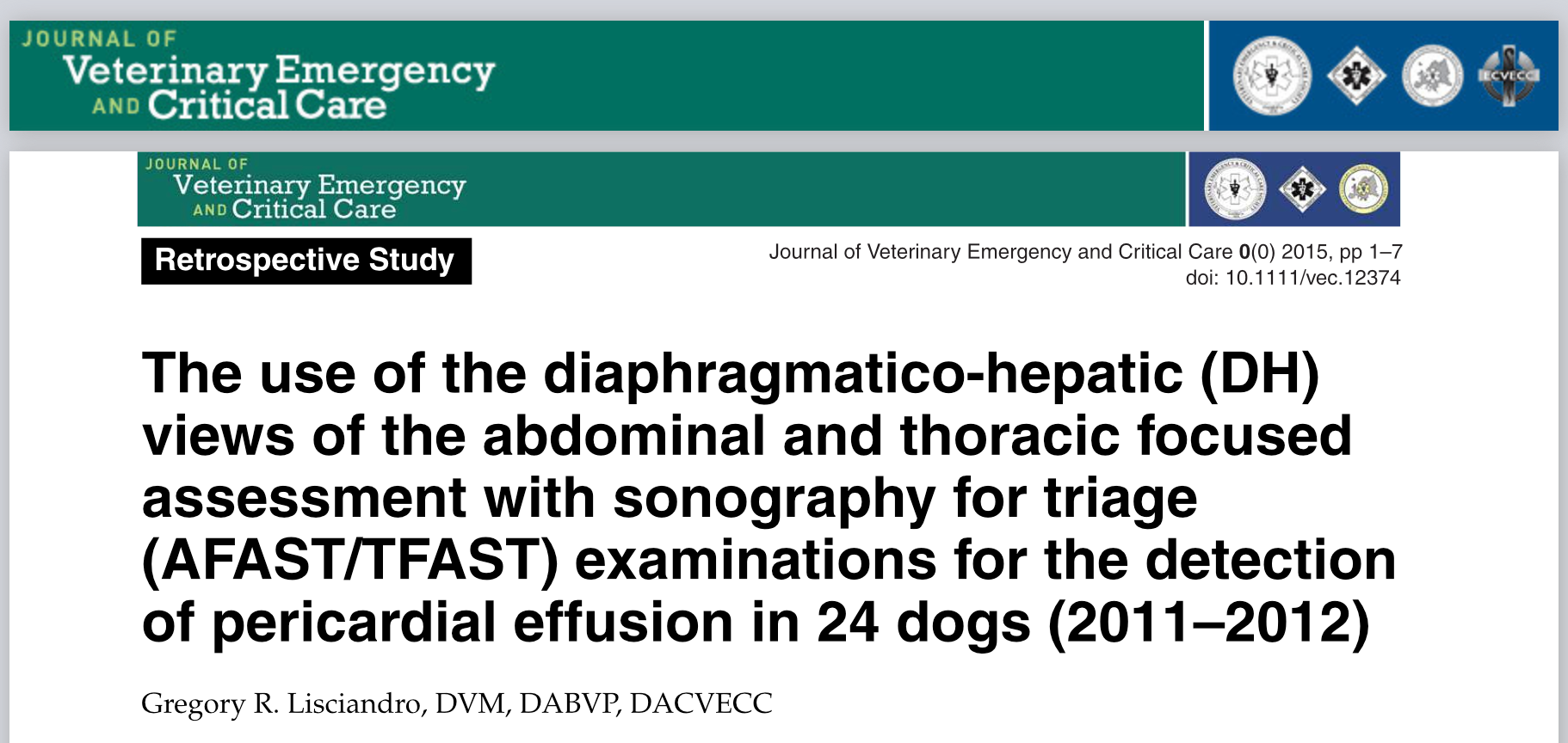
Our publication documenting how the FAST DH View serves as a rapid and reliable means to diagnose Pericardial Effusion in dogs.
Click on Image to Make Larger
More Information Regarding This Month’s ECC Blog
To obtain more information regarding the content of this month’s ECC Blog, 1) listen to our Webinars on the Gallbladder Halo Sign and the Diagnosis of Pericardial Effusion; 2) or read our textbook Point of Care Ultrasound Techniques for the Small Animal Practitioner; Wiley Publishers 2021; 3) or take our Online AFAST Course; 4) or come to our FASTVet Academy in Austin, Texas for Hands-on Training! Sign Up Now!
Click on Image to Make Larger
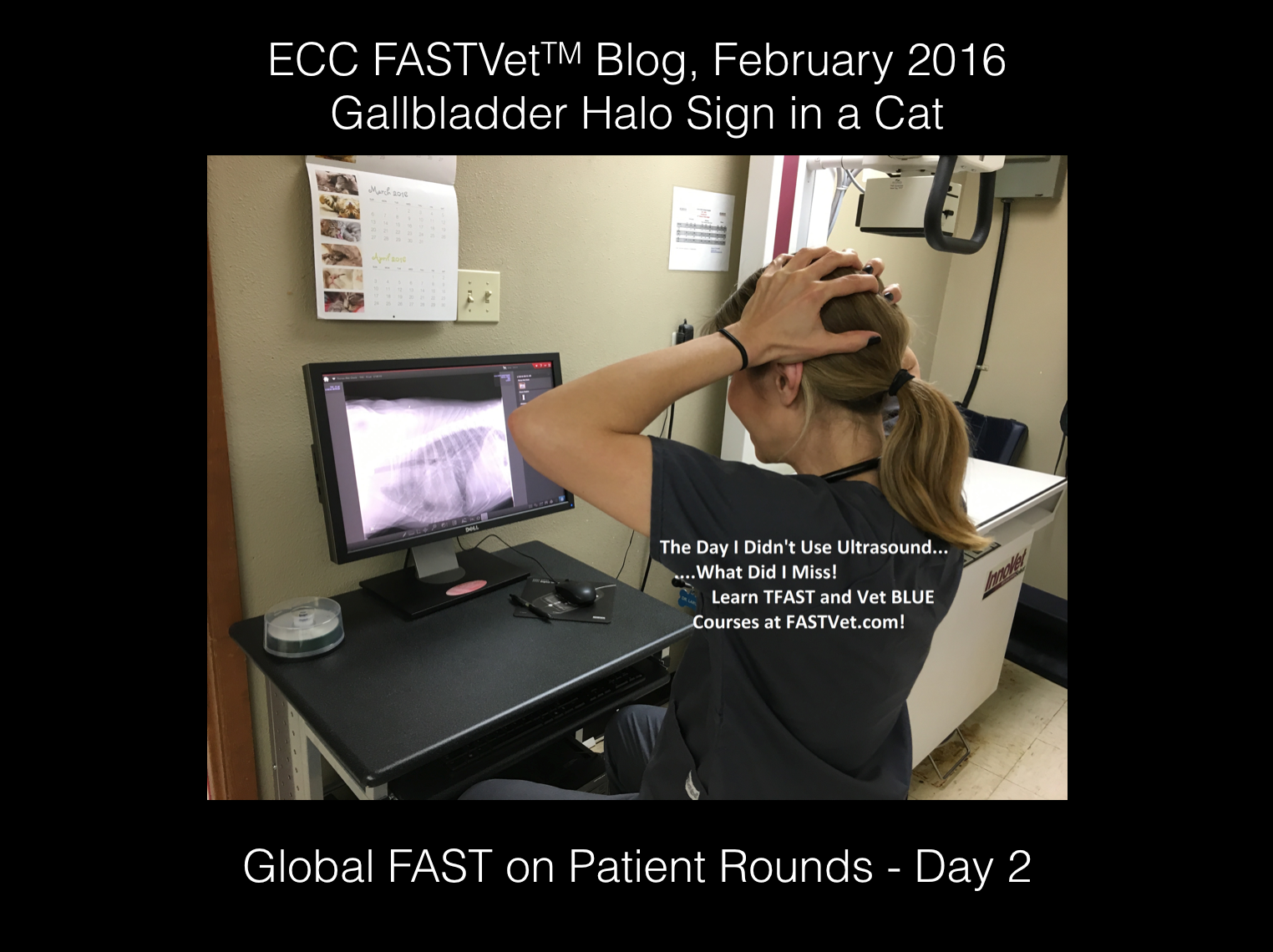
Don’t Be Like This! The Day I Did NOT Use Ultrasound! What Did I Miss!