The objective of the study was to evaluate different probes for Vet BLUE® lung ultrasound. The study design was something that Dr. Jessica Ward and I had discussed several years ago. The clinical importance and relevance was founded on the premise that many veterinary cardiologists would be tempted to use the phased-array probe for Vet BLUE® lung ultrasound evaluation immediately after or before their echocardiogram evaluation. Thus, the clinical question would be – Is the phased-array cardiac probe appropriate for Vet BLUE®?
There are 2 Major Fundamental Principles for the Probe Type (Transducer Type) to Accurately Perform Vet BLUE®:
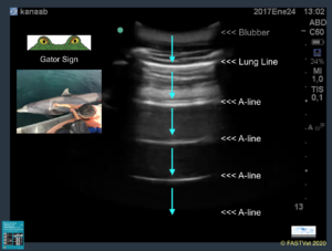
- The probe must accurately identify the “Gator Sign Orientation.” The “Gator Sign” term was published in Vet Radiol and Ultrasound in 2014 (Lisciandro, Fosgate and Fulton). The “Gator Sign” identifies the intercostal space interposed by a rib cranially and a rib caudally. The image created when the scanning plane is perpendicular to the long axis of the ribs is likened to a partially submerged alligator (Gator) peering at you.
- The probe must accurately count numbers of B-lines also called “Lung Rockets.” Our Vet BLUE® B-line Scoring System was first published in Vet Radiol and Ultrasound in 2014 (Lisciandro, Fosgate and Fulton). The highest number over a single intercostal space at each respective Vet BLUE® view is recorded and subdivided as – “Weak Positives” are “1”, “2”, and “3” and “Strong Positives” are “>3” and “♾️ (infinity using “&” on the keyboard)” using a simple easily remembered numerical system.
“Gator Sign Orientation”
The Vet BLUE® B-line Scoring System
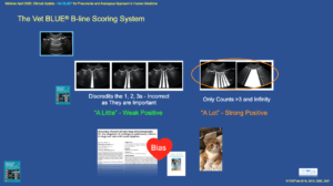
The Vet BLUE® B-line Scoring System is numerically based (“1”, “2”, “3”, “>3”, and “♾️ infinity”) as the highest number of B-lines also called “Lung Rockets” over a single intercostal space at each representative Vet BLUE® view and is composed of “Weak Positives” and “Strong Positives.”
The “Volpicelli Strong Positive Model” used in people (“>3 and infinity”) was used for the rapid screening of left-sided congestive heart failure (L-CHF) in dogs and cats, first published in J Am Vet Med Assoc in 2017 (Ward, Lisciandro, Ware et al.). However, it is important to know that “Weak Positives” are not expected during Vet BLUE® using our methodology, with the expectation in both adult dogs, adult cats, and puppies and kittens over 6-weeks of age, to have predominately “Dry Lung All Vet BLUE® Views” demonstrated in several of our clinical studies. A list of our Publications and LINKS to Pubmed and Open Access is available by clicking here.
Learn Vet BLUE® in our RACE-approved Online Courses (8-hours) with more information here.
The use of Vet BLUE® is cutting-edge and has clear advantages over other lung ultrasound formats being uniquely regional, pattern-based, with the Vet BLUE® B-line Scoring System, Vet BLUE® Severity Score, and the simple easy to learn Vet BLUE® Visual Lung Language for signs of consolidation – Shred Sign, Tissue Sign, Nodule Sign, and Wedge Sign. We were the first in the world to use proactive lung ultrasound on a daily basis having begun this paradigm change in 2005 with TFAST® lung ultrasound followed by our better screening test of Vet BLUE® (2010). Click here for more FASTVet Online Course information including Vet BLUE®.
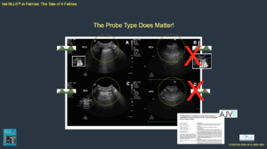
Furthermore, when viewing ultrasound images the probe type is may be determined by looking at the shape of the near field (arrows below on the image). A phased-array “cardiac” probe is a sharp point, a convex probe has a curve, and a linear probe is squared off.
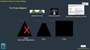
This slide is from our 9-hour RACE-approved Global FAST® Course that may be taken remotely or in-person. Note the near field of each shape representing different probe types. Click here for Global FAST® In Person Course information. We have training centers in Austin, Texas and Nashville, TN, but also travel to practices in the USA and internationally (14+ countries).
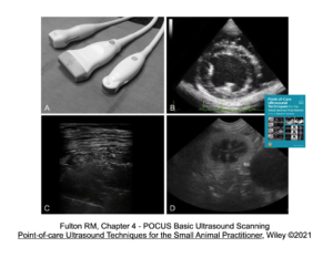
The image below is from our textbook, Point-of-care Ultrasound Techniques for the Small Animal Practitioner, 2nd edition, Wiley-Blackwell, ©2021. Click here for the textbook available off Amazon.
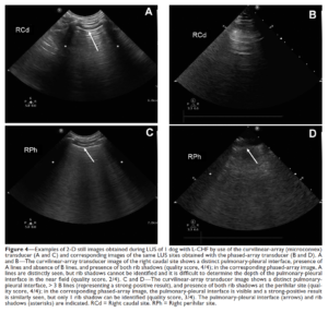
So let’s look at some images from our Am Jr of Vet Research study published in 2021 (Ward, Murphy and Lisciandro, et al. 2021). Ask the following 3 questions – 1) What probe is being used by looking in the near field of each image? and 2) Can you accurately identify the “Gator Sign Orientation”? and 3) Can you accurately count B-lines also called “Lung Rockets”?
Example 1
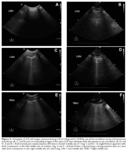
Example 2
So let’s look at some images from our Am Jr of Vet Research study published in 2021 (Ward, Murphy and Lisciandro, et al. 2021). Ask the following 3 questions – 1) What probe is being used by looking in the near field of each image? and 2) Can you accurately identify the “Gator Sign Orientation”? and 3) Can you accurately count B-lines also called “Lung Rockets”?
Example 3
So let’s look at some images from our Am Jr of Vet Research study published in 2021 (Ward, Murphy and Lisciandro, et al. 2021). Ask the following 3 questions – 1) What probe is being used by looking in the near field of each image? and 2) Can you accurately identify the “Gator Sign Orientation”? and 3) Can you accurately count B-lines also called “Lung Rockets”?
This is a slide from our all day 9-hour RACE-approved Global FAST® Course in Austin, Texas, or can be taken as our Hybrid Global FAST® Course with Online (7-hours) and Hands-on 4-hours for an 11-hour RACE-approved Global FAST® Course at our training center in Nashville, TN or we can travel to your practice and teach you on your own machine(s) – contact Dr. Greg Lisciandro for onsite training – email or text/call 210-260-5576.
The conclusion of our AJVR study was that “experience can overcome the deficiencies of the phased-array probe”; however, I am not so confident with this conclusions by my co-authors and our FASTVet recommendation would be to maximum Vet BLUE® success and use a microconvex probe that is effective for not only Vet BLUE® but for all of the Global FAST® examination. The linear probe is also acceptable and provides outstanding detail of the lung surface, but cannot be used for the entire Global FAST® examination and is better left for a radiologist’s evaluation if and when needed, rather than a sonographer performing Vet BLUE® as a screening test. This same mindset is the consensus in human point-of-care ultrasound.
Another study from Poland studied more Vet BLUE® categorized findings including its signs of consolidation, and concluded as I did, avoid the cardiac phased-array probe, its spatial resolution is not constantly detailed enough.

Send any comments regarding this FASTVet Awesome Blog to Dr. Greg Lisciandro, DVM, DABVP, DACVECC at LearnGlobalFAST@gmail.com. Become a FASTVet Premium Member and use our any resources to continue your learning!
gl/GL 4-9-2024, 1-13-2025, 1-9-2026