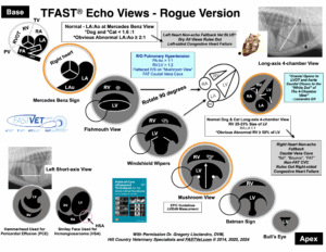
Our FASTVet TFAST® Echocardiography, Chart Updated 2025. TFAST® Echo Chart – PRINTABLE PDF version is found here. Note we teach with the reversal of the orientation consistent with all other imaging with cranial to the left and caudal to the right, thus these images are opposite of a cardiologist’s.
The Global FAST® Non-echo Fallback Views is another FASTVet Original, detailed in the 1st edition of our textbook. The principle is important for integrating information when interrelating echocardiography findings, and for the patient in which echocardiography is not possible due to patient status or need of physical and or chemical restraint that is not possible initially.
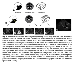
The figure above is taken from a recent Vet Clinics of North America review article by Lisciandro GR, ©2020 but is also explained well in our 2nd edition of Point-of-care Ultrasound Techniques for the Small Animal Practitioner, ©2021.
Briefly, the principles of our Global FAST® Non-echo Fallback ViewApproach…
LEFT-sided Heart Disease – Short-axis Mercedes Benz View for the LA:Ao Ratio for Left Atrial (LA) Enlargement – The strategy is that if the sonographer feels there is evidence for left atrial enlargement, thinking that left-sided congestive heart failure (L-CHF) may be present, then the sonographer integrates Vet BLUE® findings of “Wet Lung” versus “Dry Lung.” If Vet BLUE® is “Dry Lung ALL Vet BLUE® Views”, then there may be left-sided heart disease, but there is no evidence for L-CHF. This is accomplished in < 60-90 seconds point-of-care. On the other hand, if the patient has “Wet Lung” and the unique novel Vet BLUE® regional, pattern-based approach supports L-CHF over say pneumonia based on distribution of “Wet Lung”, then Vet BLUE® Severity Score can help dictate use of loop diuretics. When there is “Dry Lung” all Vet BLUE® views, or minimally “Wet Lung”, then fundamental TFAST echocardiography can wait until the patient is more stable.
If the patient has “Dry Lung”, and its fundamental TFAST® echocardiography and the application of EPIC Guidelines for declaring Stage B2 are not affirmative, then recheck the patient as per clinical judgment. See our Blog and Webinar on the EPIC Guidelines and their application during fundamental TFAST® echocardiography.
RIGHT-sided Heart Disease – Long-axis 4-chamber View for the RV:LV for Right Ventricular (RV) Enlargement – The strategy is that if the sonographer feels there is evidence for right-sided congestive heart failure (R-CHF) by enlargement of the RV, then characterization of the caudal vena cava (CVC) and its associated hepatic veins (presence or absence of the “Tree Trunk Sign”) at the AFAST®-TFAST® Diaphragmatico-Hepatic (DH) View is integrated for patient assessment. If the CVC has a “Bounce” or is “flat”, a small maximum height, then R-CHF is unlikely. There may be right-sided heart disease, but there is no evidence for R-CHF. When there is an unremarkable characterization of the CVC and its associated hepatic veins (no “Tree Trunk Sign”), then fundamental TFAST® echocardiography can wait until the patient is more stable.
Conversely, if the CVC is “FAT” (increased maximum height) especially with distended hepatic veins (positive “Tree Trunk Sign”), these findings supports (Chou et al. PLOS ONE 2021) R-CHF and a work-up is emergently needed along with R-CHF therapy. In the event that the patient has findings that are inconclusive, then the clinical should recheck the patient as per clinical judgment.
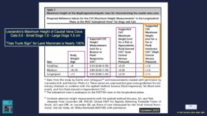
Our Table for Maximum Heights of the Caudal Vena Cava (CVC) at the AFAST®-TFAST® Diaphragmatico-Hepatic (DH) View help characterize and interpret CVC findings in addition to “Bounce”, “flat”, and “FAT.” New information, cats should never be over 0.5 cm as their CVC maximum height. Thus, the FASTVet- Lisciandro Rule for the CVC Maximum Height Measurement is now the following – “CVC Maximum Height Rule for “FAT” is <0.5 cm cats, <1.0 cm small dogs, <1.5 cm for large dogs”; thus, values over these measurements would be considered “FAT” or “Distended” or “Fluid Intolerant.” Moreover, hepatic veins positive for the “Tree Trunk Sign” is always R-CHF/right-sided volume overload until proven otherwise.
In summary, the Global FAST® Non-echo Fallback Views (Lisciandro, Editor, Point-of-care Ultrasound Techniques for the Small Animal Practitioner, ©2014, 2021; Lisciandro, J Vet Emerg Crit Care 2o11) provide important information when fundamental TFAST® echocardiography (and that by a cardiologist) is not possible because of patient status, i.e., patient instability, patient needs forms of restraint (physical/chemical). For example, “Dry Lung ALL Vet BLUE® Views” rules out L-CHF and the TFAST® echocardiography (or complete echocardiography) can wait until the patient is more stable/tolerant of physical/chemical restraint.
Moreover, when there is “Dry Lung ALL Vet BLUE® Views”, empiric loop diuretic therapy is not indicated for pulmonary edema because no clinically relevant alveolar-interstitial edema exists. If you still question your finding of “Dry Lung ALL Vet BLUE® Views”, then REPEAT the Global FAST®-Vet BLUE® in 2 to 4 to 6-hours, or during the next patient rounds, or the next recheck exam.
A “Bounce” or “flat” CVC rules out R-CHF and fundamental TFAST® echocardiography (or complete echocardiography) can wait until the patient is more stable/tolerant of physical/chemical restraint. If you still question your finding of “Bounce” or “flat” CVC, then REPEAT the Global FAST®-AFAST®-TFAST® DH View in 2 to 4 to 6-hours, or during the next patient rounds, or the next recheck exam.
We hope you found this FASTVet Blog helpful. Please send any comments to Dr. Greg Lisciandro, DVM, DAVP, DACVECC at FastSavesLives@gmail.com. Become a FASTVet Premium Member to use our many resources, and consider taking your In-Person Global FAST® Course in Austin, Texas or at your practice, and Online RACE-approved AFAST®, TFAST® and Vet BLUE®-Global FAST® Courses.
gl/GL 1-23-2025