Here are some FREE Resource for understanding Vet BLUE® lung ultrasound examination and its unique regional, pattern-based approach for a rapid respiratory working diagnosis with an easily understandable lung ultrasound signs system of “Wet Lung” vs. “Dry Lung”, its B-line Scoring System for inherent severity, its visual lung language for signs of consolidation, Shred, Tissue, Nodule and Wedge Sign, developed by the author. This system can easily be recorded and tracked in patients.
Here are some sample didactic FASTVet Webinar Shorts. Many more are available from our website and membership FASTVet.com
Ettinger 9th Edition Webinar Shorts – 02 Vet BLUE® Its Acronym and How to Perform Its Views
In this complimentary FASTVet Webinar Short Vet BLUE® the impact of “Dry Lung All Vet BLUE® Views” is covered.
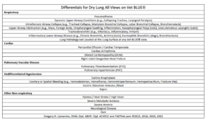
Table. Differentials for “Dry Lung All Vet BLUE® Views.” “Dry Lung” is defined as A-lines with “Lung Sliding.” “Wet Lung” is defined as B-lines with hyperechoic laser like vertical streaks that obliterate A-lines while swinging like a pendulum in respirophasic synchrony. If they don’t follow this rule, then they are not B-lines also called “Lung Rockets.” This material is reproduced with permission of John Wiley & Sons, Inc., Point-of-Care Ultrasound Techniques for the Small Animal Practitioner, 2nd Edition, Wiley ©2021 and Greg Lisciandro, Hill Country Veterinary Specialists, FASTVet.com © 2014, 2021.
Our veterinary Vet BLUE® B-line Scoring System is another FASTVet Original. Vet BLUE® B-lines are divided into a “Weak Positive” and “Strong Positive” model. We expect rare single B-lines (also called “Lung Rockets”) in adult cats and adult dogs and puppies and kittens over 6-weeks of age as documented in our published clinical research. This is important because any number of B-lines should be placed into clinical context. As per Dr. Nilam Soni, MD, at the POCUS Workshop for human medicine (Austin, Texas, January 2024), “veterinarians probably have a better B-line scoring system (than we).” Our FASTVet Vet BLUE® B-line scoring system also has advantages in recording and comparing findings to future and past studies, and has an inherent severity scoring system – see below.
Here is another complimentary FASTVet Webinar Short covering another aspect of the Vet BLUE® B-line Scoring System.
Ettinger 9th Edition Webinar Shorts – 03 Vet BLUE® and Its B-line Scoring and Weak Positives Count!
Vet BLUE® B-line Scoring System. Our Vet BLUE® B-line Scoring System taking the maximum number of B-lines over a single intercostal space at each respective Vet BLUE® region and scoring as “0”, dry, and “1”, “2”, “3”, “>3”, and “infinity” as wet. The positive B-lines as further categorized as “weak” (1,2,3) and “strong” positives (>3 and infinity). This material is reproduced with permission of John Wiley & Sons, Inc., Point-of-Care Ultrasound Techniques for the Small Animal Practitioner, 2nd Edition, Wiley ©2021 and Greg Lisciandro, Hill Country Veterinary Specialists, FASTVet.com © 2014, 2021.
Here is another complimentary FASTVet Webinar Short covering another aspect – “Pseudo B-lines” versus “True B-lines.
B-lines also called “Lung Rockets” most often represent “Wet Lung” however, there are mimics that cast the same vertical artifact but are NOT a fluid substrate cuffed by air but rather soft tissue cuffed by air, Nodule Pseudo B-line, or a semi-solid (food) in the stomach cuffed by air, Gastric Pseudo B-line. We believe it is best to call “Wet Lung” artifacts, water (CPE, NCPE), blood, or pus (pneumonia),”True B-lines” and those caused by something other than a fluid substrate, “Pseudo B-lines.” A 3rd cause of “Pseudo B-lines” would be atelectic lung.
The Probe Type Matters. Best to use a microconvex or linear probe and we do not recommend the use of a phased-array (cardiac) probe as you will see from this Blog based on a study we published in AJVR.
Algorithm. Integration of Vet BLUE® and TFAST® Echocardiography and Caudal Vena Cava. The algorithm shows integration of TFAST® echocardiography and caudal vena cava findings when B-lines are present during Vet BLUE®. This material is reproduced and modified with permission of John Wiley & Sons, Inc., Point-of-Care Ultrasound Techniques for the Small Animal Practitioner, Wiley ©2014, ©2021 and Greg Lisciandro, Hill Country Veterinary Specialists and FASTVet.com © 2021.
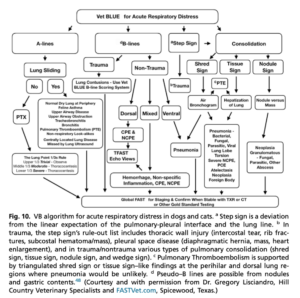
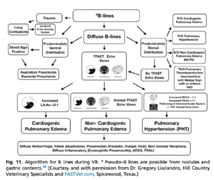
Algorithm. Vet BLUE® Signs and their Distribution for a Working Diagnosis. The algorithm shows the various Vet BLUE® findings and rule outs. This material is reproduced and modified with permission of John Wiley & Sons, Inc., Point-of-Care Ultrasound Techniques for the Small Animal Practitioner, Wiley ©2014, ©2021 and Greg Lisciandro, Hill Country Veterinary Specialists and FASTVet.com © 2021.
Some FASTVetTM Webinars – Some Require FASTVet Membership
- Vet BLUE® and TFAST® for Common Respiratory Conditions in Cats – ACVIM Forum 2022 here.
- Use of Vet BLUE® for Small Animal Pneumonia and Relevance to COVID-19 – ACVIM Forum 2022 here.
- Update TFAST® for Pulmonary Hypertension – Pearls and Pitfalls here.
- Left Atrial Tear (Rupture) – A Complication You Will See here.
- TFAST® Echo Views Pearls and Pitfalls here.
- The Tale of 4 Felines – Use of TFAST® and Vet BLUE® here.
Clinical Studies by the Author
*A List of all our 28+ peer-reviewed clinical studies may be found here.
gl/GL 4-9-2024, 1-9-2026