Case Study: AFAST® and Complete Abdominal Ultrasound (AUS) Findings in Peritonitis by Stephanie Lisciandro, DVM, Dipl. ACVIM (SAIM)
An 8-year old F/S Cocker Spaniel presented for a 5-day history of vomiting, diarrhea and inappetence. She had been seen 2-days earlier and treated empirically for non-specific gastroenteritis with Cerenia® and bland diet. On presentation, she had a fever of 104.2 and was tense but not overtly painful on abdominal palpation. CBC showed mild anemia (HCT 32%) and thrombocytopenia (52,000). Chemistry profile revealed elevation of ALT (235 U/L), ALP (982 U/L), GGT (24 U/L) and total bilirubin (1.2 mg/dl). The cPL was normal. Abdominal radiographs (AXR) showed splenomegaly and loss of serosal detail in the cranial abdomen. Initial AFAST exam was negative for free fluid. She was admitted to the hospital for supportive care. During hospitalization, she became hypoglycemic (blood glucose 52 mg/dl) but she responded to administration of intravenous dextrose. A complete detailed abdominal ultrasound (AUS) was requested.
A complete abdominal ultrasound (AUS) was performed. A second AFAST® was performed prior to the AUS examination. AFAST® showed the presence of echogenic fluid in the abdomen (abdominal fluid score 3/4 at the DH, CC and SIU views – see previous Blog on the AFAST®-applied Abdominal Fluid Scoring system here). This AFAST®-detected fluid on serial exam should have been sampled and tested and decisions made for an exploratory without the AUS. In addition, there was evidence of “lung rockets” at the Vet BLUE® caudodorsal lung regions that was also noted at the DH view along the pulmonary-pleural interface along the diaphragm.
The complete abdominal ultrasound (AUS) revealed several abnormal findings:
- There was a large volume of peritoneal effusion noted. The effusion was highly echogenic and may have been challenging to see on the initial AFAST® (see images) or very likely in dehydrated patients free fluid is not present because the patient is absorbing as much as possible to maintain intravascular volume and adhesions are “sticking” to the perforation. Echogenic fluid is consistent with cellularity, however, fluid should always be sampled and tested because ultrasound cannot reliably characterize the type of free fluid.
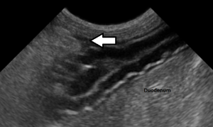
- The duodenum was corrugated and thickened (see image below).
- The mesenteric and omental fat was hyperechoic (bright white) and irregular which was consistent with steatitis.
Overt pancreatitis was not noted. - Read this important Blog on following post-op surgical cases and AUS findings here.

Fig. 1: The “free fluid” is very hyperechoic (represented by the arrows) to the adjacent liver. Echogenic fluid is consistent with cellularity however, it must be sampled and tested (PCV, film for cytology, +/- chemistries, i.e., glucose level, creatinine level, lipase level dependent on the working diagnosis).
Generally, “free fluid” is anechoic (black) in appearance. However, in this case, the fluid is highly echogenic indicating cellularity. The fluid is hyperechoic (or brighter than) to the liver and may be difficult to identify as “free fluid” to the novice ultrasonographer. The fluid is seen as the brighter bands indicated by the white arrows. However, the “free fluid” may have been in very small pockets difficult to appreciate because of patient dehydration.
The FASTVet Rule is “Rehydrate, Resuscitate, and Re-evaluate with AFAST® (always striving for Global FAST®) one more time (and continue until certain no longer needed imaging)” because of the dehydration and “sticking” of omentum to the perforation until the patient is rehydrated, resuscitated, volume restored, at which time the omentum “falls off” (no longer “sticks” to the perforation) and fluid is exuded copiously into the abdominal cavity, typically within 2-4 hours later.
Abdominocentesis was performed and should always be performed if the “free fluid” pockets are safely accessible. Grossly the fluid appeared hemorrhagic and cloudy in nature and the cellular elements of the fluid readily sedimented. Microscopic evaluation revealed a large population of neutrophils (bands and degenerate neutrophils) with a smaller population of lymphocytes and macrophages. In addition, numerous rod-shaped bacteria were seen in association with clusters of neutrophils with occasional intracellular bacteria within neutrophils and macrophages. The findings were consistent with septic peritonitis. Great images and descriptions of small intestinal normals and abnormals are in our 2nd edition textbook that may be purchased (strongly recommended as a great resource for you) through Amazon here.

Fig. 2: This image shows a “corrugated” thickened duodenum with adjacent pocket of “free fluid.” The findings are consistent with focal enteritis. Our textbook, Point-of-care Ultrasound for the Small Animal Practitioner, 2nd edition, 2021, here has great images and goes through various small intestinal findings.
There are several interesting things to discuss in this case. With regards to the peritoneal effusion, the effusion was highly echogenic. In general, “free fluid” is anechoic (black) in nature but in the case of cellular fluid, it can occasionally be so echogenic that it can be difficult to differentiate from adjacent soft tissue structures to the eyes of the less experienced ultrasonographer.
Although the definitive cause of the peritonitis could not be definitively determined based on the ultrasound alone, the fluid must be sampled and tested, the finding of a focal segment of duodenum with changes consistent with focal enteritis in addition to “free fluid” and hyperechoic fat (bright white) adjacent to the affected segment may be suggestive that the cause is related to a perforation in that segment.
Regardless, the finding of “free abdominal fluid” containing intracellular bacteria is an indication for abdominal exploratory. In this case, surgery revealed a small perforation in the proximal duodenum. It is important to note that septic peritonitis cannot be diagnosed on the basis of ultrasound alone. Cytological evaluation (sample and test) of the abdominal fluid is necessary for definitive diagnosis. In addition, septic fluid is not always as echogenic as noted in this case and may appear anechoic (black) or have variable degrees of echogenicity (grayness).
Final Comments
- This image shows a “corrugated” thickened duodenum with adjacent pocket of free fluid. The findings are consistent with focal enteritis. Another AUS image showed a “corrugated” thickened duodenum with adjacent pocket of “free fluid.” The findings are consistent with focal enteritis.
- Another interesting finding was “lung rockets” (B-lines) along the diaphragm and its deeper window into lung along the pulmonary-diaphragmatic interface. Remember from a previous Blog that the DH view is powerful for the evaluation of both the abdomen and thorax including heart, lung, pleural space for pleural effusion, and volume status through characterization of the CVC (Blog here, Webinar here). Watch this “Power of the DH View” FASTVet Webinar here.
- The presence of “lung rockets” supports pathology (alveolar-interstitial lung edema) in the caudodorsal lung regions and TFAST® with Vet BLUE® lung ultrasound is indicated for further evaluation. Thoracic radiographs (TXR) in conjunction with ultrasound are important for evaluating lung. In this case, TXR showed alveolar -interstitial infiltrates in the caudal lung lobes. In the absence of heart disease, based on the distribution and lack of left atrial enlargement on TFAST® fundamental echocardiography (Mercedes Benz view), non-cardiogenic pulmonary edema (NCPE) or inflammation consistent with acute respiratory distress syndrome was suspected. Vet BLUE® was subsequently used to follow its course and reolsution sparing the use (and cost of) of TXRs. A Webinar Short on Vet BLUE® “Wet versus Dry Lung” is found here.
gl/SCL 4-3-2025