I thought I’d toss this one out there. It’s long but this was on a radiology telemedicine service blog.
This post is truly why you part of FASTVet Nation, Global FAST are so important to bring about change in veterinary medicine!
Without biasing your response I will post my comments next week. This is posted on our Closed FaceBook Page: “FASTVet Veterinary Ultrasound that Saves Lives for Members Only” and we encourage you to join.
My Liberty to Title with My Title of this post as: “Is Dr. Wright, Dipl. ACVR, Right?”
Dr. Wright writes:
“…in the last few years, FAST (focused assessment with sonography for trauma) examinations have become popular in veterinary hospitals as a way for nonradiologists to quickly and inexpensively assess critical patients for the presence of emergent abdominal or thoracic disorders such as peritoneal or pericardial effusion.
Recently, FAST has been rebranded to FAST3 to indicate a general name for three different types of FAST scans including AFAST (abdominal fast) TFAST (thoracic fast) and Vet BLUE (bedside lung ultrasound exam). The FAST scan has essentially been “marketed” as an extension of the physical examination. FAST scans have been in use in human medicine since the 1990s.
There is a standardized protocol associated with each of these types of scans. For example, here is an introduction to the AFAST scanning protocol:
Veterinarians can learn how to FAST scan either at a conference (for example FAST scan courses are available at the yearly VECCS meeting) and online through educational websites such as Fastvet.com
So there is your 2-second introduction to FAST scanning. It is billed as an easy to learn extension of the physical examination veterinarians can do in their practice to assess critical patients.
From our vantage point, however, there might be more to this story.
Recently, we have seen a growing number of case submissions to our teleradiology service stating “No free fluid on FAST scan” while peritoneal effusion is clearly evident radiographically at the time of examination (we are seeing the radiographic images of the abdomen and not the FAST ultrasound images).
Abdominal effusion is always a concerning finding but it may be even more concerning in these cases because the clinicians may be under the false impression that peritoneal fluid is absent. Therefore, any delay in the reporting of the radiographic study could, potentially, have serious consequences for the patient. Moreover, radiographic images submitted after a negative FAST scan may be less likely to be submitted as a STAT submission because the clinician erroneously believes that there is no peritoneal effusion present.
The question we have started to ask ourselves is “what is wrong with the FAST scan?”
To get to that answer, it might help looking back at where FAST came from and what is going on in the human side. The one piece of literature that is always quoted regarding the efficacy of the FAST scan is an article that appeared in JAVMA in 2004 titled Evaluation of a focused assessment with sonography for trauma protocol to detect free abdominal fluid in dogs involved in motor vehicle accidents.In that article the authors found that in the 100 dogs in their study which presented for motor vehicle trauma, a FAST examination was found to have 96% sensitivity and 100% specificity for the detection of free abdominal fluid.
The problem with citing this study is that it may not mirror life on the ground at a veterinary hospital. This is because in the study they used “four veterinary emergency critical care (ECC) clinicians in the study. Three were residents and one was a diplomate of the ACVECC.” Granted that the specific training for the FAST scan may approximate real world conditions (“participating in this study each underwent 2 hours of didactic training in the basic physics of ultrasonography and performed FAST on 5 healthy dogs.”)
It is likely that these clinicians are far more experienced with ultrasound than many veterinarians who participate in FAST scan courses. Based on our experience working with emergency clinicians vs. referring veterinarians with no previous experience with ultrasound (and who also have less experience in treating patients with vehicular trauma) using four ECC clinicians as the cohort for the study likely introduced significant selection bias toward a successful outcome.
The knee jerk reaction to this blog post so far would be to (understandably) conclude something along the lines of “here is another radiologist just trying to protect their turf and not allow rDVM’s to touch the ultrasound probe.”
Again, if that is the response it would be reasonable. In this case, however, nothing could be farther from the truth.
The first reason you would be wrong if that was your conclusion is that we were one of the first groups of radiologists to actually recommend that there should be an ultrasound probe in every veterinary hospital.
The second reason is that we just sponsored a Vet BLUE course at a recent meeting because we are confident that FAST3 scanning techniques are (at least theoretically) beneficial.
The third reason that you would be wrong is that our opinion is influenced by new research on the human side.
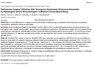
In the April 2017 edition of JACR it seems that there may be a similar trend on the human side. In an article titled, downstream imaging utilization after emergency department ultrasound interpreted by radiologists versus nonradiologists: a Medicare claims-based study there was a reduction in downstream (e.g. secondary imaging) performed when an ultrasound examination is interpreted by a radiologist rather than a nonradiologist.
This article did not go into the reasons why this might be other than to say that the “documented higher use of limited ultrasound examinations by nonradiologists or a lack of confidence of in the interpretation of nonradiologists may potentially explain this increase in follow up imaging examinations.”
Based on our observations we contend that although we believe in the concept of the FAST scan – something is wrong with the implementation in veterinary medicine given that we are seeing an increasing number of faulty FAST scans performed by referring veterinarians.
It would be irresponsible to try to guess the causality behind the increased number of faulty FAST scans that are submitted to our teleradiogy service as any number of factors could be at play including technician error when submitting the clinical history, poor education when learning to FAST scan, we are only seeing the cases submitted where the ultrasound findings were actually equivocal or complicated (e.g. positive fast scans are not submitted for follow up teleradiology so there is a selection bias for our observation), the ultrasound machines veterinarians are using are not up to the task (for example it can be extremely difficult to scan very large dogs with hand held ultrasound machines), etc.
BOTTOM LINE: we support the use of ultrasound by general practitioners. Unfortunately, we are seeing an uptick in the number of faulty FAST scan results in our teleradiology service. We do not know why and it would be irresponsible to attempt to guess why although we suspect that training is part of the problem.
The goal of this post is to serve as an early warning indicator that we are potentially seeing a problem in veterinary medicine that could result in poor patient care and/or added expense to the pet owner. If we are correct, those are two unintended consequences of FAST scanning adoption in veterinary medicine.” – End of Dr. Wright’s Blog.
P.S. Here is the Abstract on the study Dr. Wright refers – FYI. Click on it to make it larger and readable. Is there a weakness in the study design of this study? I argue there is, and its a biggie!


