This is a case presentation by Dr. Stephanie Lisciandro, DVM, DACVIM (SAIM).
“Cinnamon” is a 3 year old F/I French Lop Rabbit who presented for lethargy and decreased appetite. On initial evaluation, she had notable abdominal distention. An AFAST® was performed by the referring veterinarian and she had an abdominal fluid score of 4/4 (positive at DH, SR, CC, and HR sites). A complete abdominal ultrasound was requested.
On initial evaluation prior to the abdominal ultrasound, it was noted on physical exam that she was moderately tachypneic. Thoracic auscultation revealed a gallop rhythm with no heart murmur and bilateral crackles with an increased respiratory rate. A Global FAST®, defined as the combination of AFAST®, TFAST®, Vet BLUE as a single ultrasound examination, was recommended prior to a more detailed abdominal ultrasound and complete echocardiography because I was concerned about the rabbit’s stability. The findings were as follows:
TFAST® performed with the following findings:
- Left CTS (chest tube site): Normal “lung sliding” (no pneumothorax, PTX)
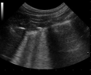
- Left CTS B lines: Present –interstitial lung fluid is evident
- Right CTS: Normal “lung sliding” (no pneumothorax, PTX)
- Right CTS B lines: Present –Interstitial lung fluid is evident
- Right PCS (pericardial site) view: Pleural effusion (PE) is noted. No pericardial effusion (PCE) is noted.
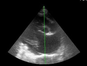
- TFAST® views of the heart showed marked left atrial enlargement, left ventricular dilation with decreased contractility.
Vet BLUE® Lung Ultrasound: A Vet BLUE® was performed with the following findings:
- Left hemithorax (CdLR, PhLR, MLR, CrLR): 0, 1, infinity, infinity; pleural effusion is noted cranially
- Right hemithorax (CdLR, PhLR, MLR, CrLR): infinity, infinity with small shreds, infinity, infinity; and pleural effusion is noted cranioventrally
 Interpretation: There is evidence of severe interstitial edema bilaterally with focal shred signs (more severe on the right side). This finding is consistent with fulminant pulmonary edema (cardiogenic or non-cardiogenic). In addition, there is moderate volume pleural effusion noted cranially on both sides, bilaterally. These Vet BLUE® findings may support cardiac disease and a complete echocardiogram is recommended.
Key – CdLR=caudal dorsal lung region, PhLR=perihilar lung region, MLR=middle lung region, CrLR=cranial lung region
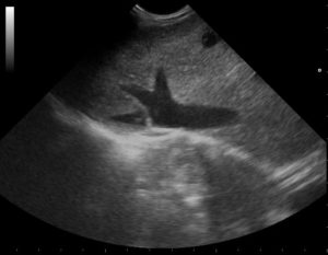
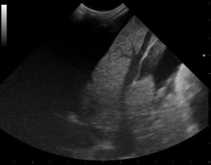
AFAST® showed large volume abdominal effusion at DH, SR, CC and SIU views with an abdominal fluid score of 4/4. Marked hepatic venous distention, the “Tree Trunk Sign” was noted at the DH views and evaluation of the caudal vena cava (CVC) showed distention, called “FAT” and “fluid intolerant”, without the expected “Bounce”, called “fluid responsive.” Hepatic veins are seen as “Tree Trunks” in the images below. The first of the three images shows marked distention of the caudal vena cava and hepatic veins at the AFAST® DH view. In the center image, in addition to marked hepatic venous distention, there is free fluid around the margins of the liver.”Free fluid” may be differentiated from vessels through the use of color flow Doppler. Key – DH=Diaphragmatico-hepatic, SR=Spleno-renal, CC=Cysto-colic; and SIU=Spleno-intestino Umbilical
The image to the far left shows the right parasternal short-axis view of the heart at the heart base called the Mercedes Benz View. This view shows marked left atrial enlargement (tear drop like shape below the aorta) when compared to the aorta (center, circle in the middle of the screen).
A complete echocardiogram was performed and showed marked left atrial enlargement (LA:Ao ratio 2.8; normal is < 1.2) as well as marked right atrial dilation. Both right- and left-sided congestive heart failure (L-CHF) was present based on the presence of pulmonary edema with marked left atrial enlargement and pleural effusion and ascites with distention of the caudal vena cava, called “FAT” or “fluid intolerant” and the associated hepatic veins, called a “Tree Trunk Sign”, another FASTVet original term from the 1st edition of our textbook that is now in the peer-reviewed veterinary literature. The “Tree Trunk Sign” is nearly 100% specific for R-CHF for dogs in a study of which I co-authored, Chou et al. PLosOne 2022. See our Publications by clicking here.
Congestive heart failure (CHF) is fairly common in domestic rabbits. In this instance, we show the importance of information that can be gained by using Global FAST® in this species, a species that is very difficult to radiographically image due to the risk of transport, and restraint (physical and chemical). Moreover, our FASTVet Global FAST Non-echo Fallback Views, other FASTVet original, provided information regarding not only the presence but also the degree left-sided CHF through the low impact rapid use of our Vet BLUE®with its B-lines soaring system (0,1,2,3,>3, infinity), and its inherent severity scoring system with B-line scoring over the Vet BLUE® regional distribution.
This rabbit was then treated with furosemide and pimobendan.
On recheck examinations Vet BLUE® and Global FAST® may be used to help guide clinical course and medications. For example, by the degree and distribution of “wet lung” using our Vet BLUE® B-line scoring system, loop diuretic dosing may be guided.
Take our Online Global FAST® Courses, Our Hands-on In-person Global FAST® Courses (we have courses in Austin and travel throughout the USA and internationally), get our 2nd Edition of our textbook Point-of-care Ultrasound Techniques for the Small Animal Practitioner that has chapters on Exotics, and become a FASTVet Premium Member to geek out and learn our cutting-edge approach.
gl/GL 11-1-2024


