What is AFAST®?
FAST is an acronym that stands for Focused Assessment with Sonography for Trauma. It is an ultrasound exam developed by trauma surgeons, yes trauma surgeons, in the 1990s and used as a screening test for the detection of free fluid in the abdominal cavity, ascites, and the pleural cavity including pleural effusion and pericardial effusion. In 2004 the translational study from humans to dogs was published by Boysen and colleagues out of Tufts.
The next year we developed AFAST® as part of my clinical research requirement for my emergency and critical care residency training out of the Emergency Pet Center, San Antonio, Texas. We made changes from the original format by modifying and renaming its views and adding a fifth umbilical view. These views were named according to their target organs to improve teaching (the sonographer knows what fundamental anatomy to expect) while making AFAST® a screening test for not only free fluid but also soft tissue abnormalities (a radical change).
Moreover, scanning planes, screen orientation, and probe manipulations were standardized to enhance the learning process. Patients were no longer shaved (a radical change). Right lateral was preferred over left lateral recumbency because in right lateral fundamental echocardiography, electrocardiography, imaging the gallbladder and caudal vena cava, and performing abdominocentesis were more amenable. However, AFAST® could also be performed in standing position being even lower impact for the patient (a radical change).
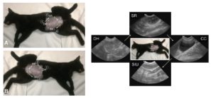
Figure. The first 4-views of AFAST® are used for abdominal fluid scoring. The Hepato-Renal view has been renamed the Spleno-Intestino Umbilical (SIU) view. Note not shown is the Hepato-Renal 5th Bonus view. The ultrasound images are examples of the starting point and the depth/relative proportionality used at each respective view regardless of mammalian species. This cat was sedated for a surgical procedure. This material is reproduced with permission of John Wiley & Sons, Inc., Point-of-Care Ultrasound Techniques for the Small Animal Practitioner, 2nd Edition, Wiley ©2021 and Greg Lisciandro, Hill Country Veterinary Specialists, FASTVet.com.
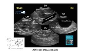
Figure. It is easy to master the fundamental AFAST® anatomy at each respective AFAST® view in longitudinal orientation making everything spatially like a lateral abdominal radiograph. You can memorize these 4 AFAST® views making this an easily mastered achievable ultrasound skill. This material is reproduced with permission of John Wiley & Sons, Inc., Point-of-Care Ultrasound Techniques for the Small Animal Practitioner, 2nd Edition, Wiley ©2021 and Greg Lisciandro, Hill Country Veterinary Specialists, FASTVet.com.
When Should You Perform AFAST®?
The mindset was to use AFAST® as “an extension of the physical exam.” In other words, everyday ultrasound nearly every patient. An AFAST® is a <3-minute test achievable with minimal ultrasound training.
Indications for an AFAST®
Initially we used AFAST® for trauma, triage, and tracking (monitoring). To avoid the onslaught of confusing acronyms in human medicine with formats having similar objectives, we added the T3 (trauma, triage, tracking) to our acronyms, i.e., AFAST3. However, we quickly morphed to the following clinical applications:
- Any abnormal or questionable patient
- Your new preanesthetic screening test
- Perioperative monitoring
- Semiannual and annual visits
- Geriatric screening test
- During patient rounds and recheck exams
AFAST® has become so widespread for these indications that we have more recently dropped the T3. The AFAST®approach allows for cost-effective imaging that otherwise would not be performed. For example, we out-price ourselves with radiography and complete abdominal ultrasound studies. Thus, many patients get sent home without any imaging. AFAST® gives you a “baby step” to screen for the need for additional imaging including radiology, complete ultrasound studies, and computed tomography (CT).
By “seeing” your problem list (evidence based information) the diagnostic plan is better streamlined and treatment more accurately determined. And, if you think about it, an AFAST® is a better initial screening imaging test than abdominal radiography for most of our patients.
Advantages of AFAST®
AFAST® is rapid, radiation sparing, low impact (minimal restraint), and real-time information (no delay) right there at your patient’s side. With proper training and minimal experience an AFAST® exam should take < 3-minutes.
AFAST® can not only determine the presence or absence of ascites but can also semi- quantitate volume by the AFAST®-applied abdominal fluid scoring system (our next blog). Moreover, many of the soft tissue abnormalities detected during AFAST® are only suspected or completely missed based on physical exam, blood and urine testing, and plain radiography.
|
AFAST® and Its Target-organ Approach for Soft Tissue Abnormalities
|
|
| Diaphragmatico-Hepatic View (DH) | Liver:
Masses, Cysts, Heterogeneous Echogenicity Gallbladder: Sediment, Sludge, Mucoceles, Wall Abnormalities, Common Bile Duct Distension Caudal Vena Cava and Hepatic Veins: Distension, Dirofilaria, Thrombi, Masses Lung: B-lines, Signs of Consolidation
|
| Spleno-Renal View (SR) | Left Kidney:
Mineralization, Calculi, Pyelectasia, Hydronephrosis, Cortical Cysts, Perinephric, Cysts, Ureteral Distension, Cortical Infarction, Masses Spleen: Masses, Heterogeneous Echogenicity (Lymphoma, Torsion) Retroperitoneal Space: Masses, Thrombi, Free Air
|
| Cysto-Colic View (CC) | Urinary Bladder:
Sediment Calculi Thrombi Masses Uterus: Fluid-filled (Pyo-, Hydro-, Muco-metra) Pregnancy Caudal Abdominal Masses
|
| Spleno-Intestino Umbilical View (SIU) | Spleen:
Masses,Heterogeneous Echogenicity (Lymphoma, Torsion) Small Intestine: Ileus and Distension Wall Abnormalities Masses Mid-abdominal Masses Gastric Distension Hepatomegaly
|
| Focused Spleen | Spleen:
Masses, Heterogeneous Echogenicity (Lymphoma, Torsion)
|
| Hepato-Renal 5th Bonus View | Right Kidney:
Mineralization, Calculi, Pyelectasia, Hydronephrosis, Cortical Cysts, Perinephric Cysts, Ureteral Distension, Cortical Infarction, Masses Liver: Masses, Cysts, Heterogeneous Echogenicity
|
| This material is reproduced and modified with permission of John Wiley & Sons, Inc., Point-of-Care Ultrasound Techniques for the Small Animal Practitioner, Wiley ©2014,2021 and Greg Lisciandro, Hill Country Veterinary Specialists and FASTVet.com ©2020
|
|
AFAST® add-on skills include characterization caudal vena cava and hepatic veins (patient volume status), the detection of pneumoperitoneum (free air), the assessment of gastrointestinal motility (ileus), and the assessment of pulsatile flow through intra-abdominal and retroperitoneal structures (viable, torsed, thrombosed).
AFAST®: Talking Points for Clients
The spiel is important to be standardized within the practice so that your clients aren’t confused. An example of a spiel would be as follows:
“We are going to perform an AFAST® ultrasound exam on your pet. AFAST® is a screening test looking for any free fluid, forms of peritonitis, within the abdominal cavity of your pet. Free fluid could be blood, pus, or something else. We also are looking for any obvious soft tissue abnormalities in your pet’s major abdominal organs.”
Optional – “If you presented at Methodist Hospital here in San Antonio, a FAST ultrasound exam would be similarly used as a first line screening test providing information point-of-care without delay that better helps lead to a diagnosis and more accurate treatment in many instances.”
For critical triaged patients – “If there is free fluid that is safely accessible, may I use a needle about the same size we vaccinate with to sample the fluid and test it.”
This quickly gives you permission to do the AFAST® and abdominocentesis when minutes count rather than going back and forth to the client for permissions. By keeping it simple, less questions will be asked so you may return and care for the patient.
AFAST®: Recording Findings
Goal-directed templates (GDTs) for recording your findings are a must for success. They keep you disciplined and on task. AFAST® is performed the same order every time just like a cardiologist and radiologist perform their ultrasound examinations the same way every time. GDTs also give you value for comparison to future exams for you and for your colleagues.
Examples of GDTs may be found at our website FASTVet.com on our Free Resources page and clicking here.
References & Further Reading
- Lisciandro GR, Lagutchik MS, Mann KA, et al. Evaluation of an abdominal fluid scoring system determined using abdominal focused assessment with sonography for trauma (AFAST) in 101 dogs with motor vehicle trauma. J Vet Emerg Crit Care 2009; 19(5):426-437.
- Lisciandro GR. Focused abdominal (AFAST) and thoracic (TFAST) focused assessment with sonography for trauma, triage and monitoring in small animals. J Vet Emerg Crit Care 2011;20(2):104-122.
- Lisciandro GR. Lisciandro GR. The use of the diaphragmatico-hepatic (DH) view of the abdominal and thoracic focused ultrasound techniques with sonography for triage (AFAST/TFAST) examinations for the detection of pericardial effusion in 24 dogs (2011-2012). J Vet Emerg Crit Care 2016;26(1):125-31.
- Lisciandro GR, Fosgate GT, Romero LA, et al. The expected frequency and amount of free abdominal fluid estimated using the abdominal FAST-applied abdominal fluid scores in healthy adult and juvenile dogs. J Vet Emerg Crit Care 2021 31(1):43-51.
- Lisciandro GR. Chapter 6: POCUS: AFAST-Introduction and Image Acquisition. In Point-of-care Ultrasound Techniques for the Small Animal Practitioner, 2nd Edition, Ed. Lisciandro GR. Wiley Blackwell: Ames IA 2021.
- Lisciandro GR. Chapter 7: POCUS: AFAST-Clinical Integration, Point-of-care Ultrasound Techniques for the Small Animal Practitioner, 2nd Edition, Wiley-Blackwell: St. Louis, ©2021.
- Lisciandro GR. Cageside Ultrasonography in the Emergency Room and Intensive Care Unit. Vet Clin North Am Small Anim Pract 2020;50(6):1445-1467.
- Lisciandro GR. Chapter 3: Point-of-Care Ultrasound. In Small Animal Diagnostic Ultrasound, 4th Edition, Eds. Mattoon JS, Sellon R, Berry CR. Elsevier: St. Louis MO, 2021.
- Chou Y, Ward JL, Baron LZ, Murphy SD, Topf MA, Lisciandro GR, et al. Focused ultrasound of the caudal vena cava in dogs with cavitary effusions or congestive heart failure: a prospective observational study. PLoS One 2021;16(5):e0252544. doi:10.1371/journal.pone.0252544
- Lisciandro GR, Gambino JM, Lisciandro SC. Case series of 13 dogs and 1 cat with ultrasonographically-detected gallbladder wall edema associated with cardiac disease. J Vet Intern Med 2021 35(3):1342-1346.