
Authors: Johnson CB, Nobles IJ, Hodges SC, Lisciandro GR
Johnson CB, Nobles IJ, Hodges SC – Oklahoma Veterinary Specialists, Tulsa, OK USA
Lisciandro GR – Hill Country Veterinary Specialists and FASTVet.com, Spicewood, TX USA
Short Version
Title: The Frequency of Aspiration Pneumonia Using the Vet BLUE® Protocol in Dogs Presenting to the Emergency Department for Vomiting or Regurgitation
Introduction: Vomiting and regurgitation are common presenting complaints in dogs evaluated in the emergency department (ED) and are risk factors for aspiration pneumonia/pneumonitis. Although a variety of risk factors for aspiration pneumonia/pneumonitis have been documented in the literature, its true prevalence in the dog is unknown. As morbidity and mortality associated with aspiration pneumonia can be high, early detection, monitoring, and treatment is important to improve outcomes. Point-of-care lung ultrasound has proven effective in rapidly identifying various types of pneumonia in people and dogs. The objective of this study was to characterize the incidence of aspiration pneumonia/pneumonitis in dogs presenting to the ED with the primary complaint of vomiting or regurgitation.
Methods: Seventeen client-owned dogs presenting for vomiting or regurgitation were evaluated at a multispecialty private practice. All patients had point-of-care ultrasound imaging performed following the Global FAST® protocol using the combination of AFAST®, TFAST®, and Vet BLUE® lung ultrasound. Free fluid in cavities or spaces and soft tissue abnormalities were recorded in addition to lung ultrasound findings supportive of aspiration pneumonia, i.e., B-lines, shred sign and tissue sign detected within gravity dependent Vet BLUE® acoustic window of its middle and cranial lung regions. Confirmative thoracic radiography was performed. Clinical signs at the time of aspiration pneumonia/pneumonitis diagnosis were recorded.
Results: Three of 17 dogs (17.6%) were found to have aspiration pneumonia/pneumonitis on lung ultrasound Vet BLUE® protocol that was confirmed with thoracic radiographic findings. None of these three dogs had clinical evidence of aspiration pneumonia, i.e., none had cough, fever or abnormal respiratory parameters, i.e., respiratory rate, pulse oximetry. None of these dogs had any free fluid detected, i.e., no pleural or pericardial effusion and no ascites.
Conclusions: Although a small case series, it appears that aspiration pneumonia/pneumonitis may appear more frequently as a silent condition in the ED and that larger case numbers are worthy of study for a true representation of prevalence with this pulmonary complication.
Long Version
Title: The Frequency of Aspiration Pneumonia Using the Vet BLUE® Protocol in Dogs Presenting to the Emergency Department for Vomiting or Regurgitation
Introduction:
Vomiting and regurgitation are common presenting complaints in dogs evaluated in the emergency department (ED) and are risk factors for aspiration pneumonia/pneumonitis. Although a variety of risk factors for aspiration pneumonia/pneumonitis have been documented in the literature, its true prevalence in the dog is unknown. As morbidity and mortality associated with aspiration pneumonia can be high, early detection, monitoring and treatment are important to improve outcomes. Dogs with aspiration pneumonia/pneumonitis can have a wide array of clinical signs, physical exam findings and clinicopathological abnormalities. Aspiration pneumonia can be difficult to diagnose in the canine patient and is often a diagnosis of presumption. With the clinical presentations associated with aspiration pneumonia being sometimes nebulous, new techniques to improve detection are essential. Point-of-care lung ultrasound has proven effective in rapidly identifying various types of pneumonia in people and dogs. The objective of this study was to characterize the incidence of aspiration pneumonia/pneumonitis in dogs presenting to the ED with the primary complaint of vomiting or regurgitation.
Methods:
Seventeen client-owned dogs presenting for vomiting or regurgitation were evaluated at a multispecialty private practice. All patients had point-of-care ultrasound imaging performed by a non-blinded emergency clinician trained to perform Vet BLUE® following the Global FAST® protocol using the combination of AFAST®, TFAST® and Vet BLUE® lung ultrasound. Free fluid in cavities or spaces and soft tissue abnormalities were recorded in addition to lung ultrasound findings supportive of aspiration pneumonia, i.e., B-lines, shred sign and tissue sign detected within gravity dependent Vet BLUE® acoustic windows of its middle and cranial lung regions. Confirmative thoracic radiography was performed. Clinical signs at the time of aspiration pneumonia/pneumonitis diagnosis were recorded. Aspiration pneumonia was defined as persistent pulmonary consolidation with dynamic air bronchograms in one or multiple lung fields using Vet BLUE® with or without clinical signs of fever, cough, tachypnea, or dyspnea.
Results:
Three of 17 dogs (17.6%) were found to have aspiration pneumonia/pneumonitis on lung ultrasound that was confirmed with thoracic radiographic findings. None of these three dogs had clinical evidence of aspiration pneumonia, i.e., none had cough, fever or abnormal respiratory parameters, i.e., respiratory rate, pulse oximetry. None of these dogs had any free fluid detected, i.e., no pleural or pericardial effusion and no ascites.
Conclusions:
Using a pattern based approach to detect pulmonary consolidation, Vet BLUE® can rapidly and effectively aid in the diagnosis of aspiration pneumonia/pneumonitis. The routine use of Vet BLUE® on canine patients presenting with vomiting and/or regurgitation can aid in the earlier diagnosis and therefore intervention of patients with presumptive aspiration pneumonia/pneumonitis. Further investigation should be pursued with larger case numbers for a more accurate representation of the frequency associated with this pulmonary complication.
Supplementary Figures
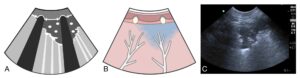
Shred Sign – Air Bronchograms on Lung Ultrasound. The hallmarks of the Shred Sign are the bright white (hyperechoic) punctate areas within the lung consolidation representing air bronchograms. The Vet BLUE® regional pattern-based approach is always used to determine where lung ultrasound findings are located for your working diagnosis. Gravity dependent asymmetrical distribution is the hallmark of aspiration and bacterial bronchopneumonias. ©Gregory Lisciandro, FASTVet.com 2014-2023.

The Vet BLUE® Visual Lung Language of Dry Lung, Wet Lung (B-lines and lung rockets), and consolidation when now enough disease has displaced air that the echoes can penetrate into the lung parenchyma – Shred Sign (air bronchograms), Tissue Sign (hepatization of lung), Nodule Sign, Wedge Sign (supports PTE when in non-gravity dependent upper regional views of caudodorsal and perihilar). ©Gregory Lisciandro, FASTVet.com 2014-2023.
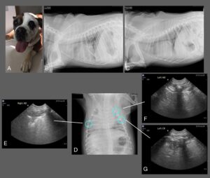
An example of aspiration pneumonia detected on Vet BLUE®. Note the difficulty in interpreting the thoracic radiograph that is clear on Vet BLUE® with bilaterally detected Shred Signs performed more rapidly with minimal to no patient restraint and without risk of transport and restraint of radiography. ©Gregory Lisciandro, FASTVet.com 2014-2023.
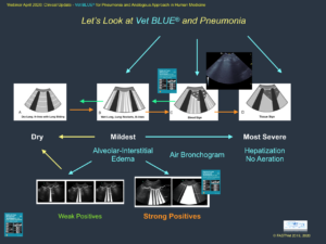
Our FASTVet Original for tracking aspiration pneumonia using Vet BLUE® following that is mildest with B-lines or lung rockets, to more severe with Shred Sign, to most severe with Tissue Sign. Moving right is worsening and moving left is improving to resolution of Dry Lung all Vet BLUE® views. ©Gregory Lisciandro, FASTVet.com 2014-2023.