Special thank you to my co-authors – Drs. Aubrey Hnatusko, DACVECC and John Gicking, DACVECC.
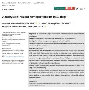
This case series documents 11 dogs with confirmed anaphylactic hemoabdomen first described by Lisciandro GR in 2016 published as an Abstract in J Vet Emerg Crit Care.

Some important points:
- All 11/11 (100%) dogs presented with acute weakness or collapse
- Vomiting and hematochezia were also present in 6/11 (55%)
- None (0%) had any cutaneous signs indicative of a hypersensitivity reaction
- None (0%) had a witnessed envenomation, i.e. witnessed bee sting
- All had hemoabdomen confirmed by abdominocentesis and an abdominal packed cell volume (PCV) compared to a peripheral venous PCV of ≥50%
- All (100%) had sonographic striation of the gallbladder wall, the so-called “gallbladder halo sign”
- AFAST®-assigned fluid scores were as follows:
- small volume bleeders (27%) as AFS 1 (2/11); AFS 2 (1/11) for a total of 3/11
- large volume bleeders (73%) as AFS 3 (4/11) and AFS 4 (4/11) for a total of 8/11
- The frequency of Positive AFAST Views was as follows:
- DH (82%, 9/11)
- SR (64%, 7/11)
- CC (73%, 8/11)
- HR (64%, 7/11)
- TFAST® showed no pleural or pericardial effusion (0%) in the 5/11 dogs examined
- *expect no other sites of cavitary bleeding
- #note 5/11 dogs had Global FAST performed
- Vet BLUE® showed Dry Lung All Views (0%) in the 5/11 dogs examined
- *expect no alveolar-interstitial pulmonary edema in single insult anaphylaxis
- #note 5/11 dogs had Global FAST performed
- All survivors (10/11) survived without surgical intervention being treated medically. See our FASTVet Treatment Sheet by clicking here
*Also based on having treated over 100 canine medically-treated anaphylactic hemoabdomen
#Global FAST® prevents imaging interpretation errors and searches for additional complications and always is recommended
Listen to the MUST WATCH – Case-based Webinar on Anaphylactic Hemabdomen by clicking here
The Abstract may be found on PubMed by clicking here
