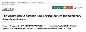
Introduction – Wedge Sign
We have known that Vet BLUE® could help screen for pulmonary thromboembolism (PTE)/pulmonary embolism (PE) for over a decade by finding the “Wedge Sign” – our created term. Just like a renal infarction, lung infarction is the same, a triangular or wedge shape of necrosis in its most extreme periphery. The Wedge Sign is more of a Tissue Sign than a Shred Sign and is indistinguishable from pneumonia/other pathology of consolidation when in gravity-dependent Vet BLUE® regions. However, when the Wedge Sign is in upper non-gravity dependent Vet BLUE® regions of caudodorsal and perihilar where pneumonia is not expected, PTE/PE should be suspected and listed as a differential.
Differentiating PTE from PE
PTE is when the thrombus if formed away from the lung, breaks off, and travels to lung causing infarction. PE is when the organ of the thrombus is directly at the site of lung infarction.
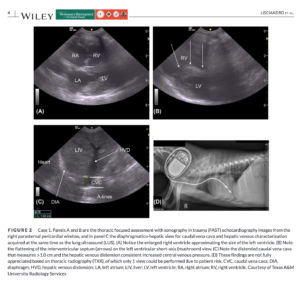
Use of TFAST® and Global FAST®
Integrating other information during Global FAST® is important to look for any sonographic risk factors and co-morbidities. TFAST® is used for several reasons including acute right ventricular dilation, distended pulmonary arteries, thrombi including saddle thrombus at the bifurcation of the pulmonary arteries at or just above the Mercedes Benz view on short-axis and on long axis, and detection of heartworms (Dirofilaria immitis).
During AFAST®, ascites and soft tissue abnormalities such as intra-abdominal masses including lymph nodes, torsions, renal infraction, etc. may be detected and placed into clinical context.
Our Publication
A big Texas thank you to my co-authors, Drs. Melanie Puchot, DACVIM (SAIM) and Stephanie Lisciandro (my wife), DACVIM (SAIM) both specialists in small animal internal medicine (SAIM), and Dr. Jen Gambino, DACVR. We submitted 3 case examples, 2 of which were accepted by the reviewers. We will outline the cases below and some of the highlights of each. The article is in the Journal of Veterinary Emergency and Critical Care and available in Early View at the time of this blog and has been listed on PubMed.
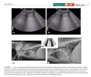
Case 1
The dog had several risk factors, a pacemaker lead, a Urine Protein:Creatine Ratio (UPC) of 15, and recent prednisone administration for its collapsing trachea. Our published Figures should be explanatory.
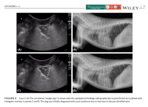
Case 2
The dog had as a major risk factor, heartworm (Dirofilariasis) disease with caval syndrome. Our published Figure should be explanatory.
We encourage you to become a FASTVet Premium Member and listen to our Pulmonary Hypertension Webinar that also includes managing caval syndrome. Please send any comments or suggestions to Dr. Greg Lisciandro, DACVECC at LearnGlobalFAST@gmail.com
We hope you enjoyed and learned from this FASTVet Blog.