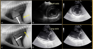
The TFAST® Views of the heart are easily learned with basic principles in mind during cardiac evaluation. Moreover, the gestalt or eyeball method has gained momentum as a reliable means to assess cardiac function without the technical challenges and lengthy time involved with M-mode and color flow Doppler (clinical research by Paula Ferrada, MD).
What are the basic TFAST® Views of the Heart?
- The Left Ventricular (LV) Short-axis View (LVSAV, the so called “mushroom” view)
- The Left Atrial to Aortic Ratio (LA:Ao, the so called “quick peek” view on short-axis)
- The Right Ventricle to Left Ventricle Ratio (RV:LV on long-axis)
You may click on the image to enlarge it.
From Focused Ultrasound Techniques for the Small Animal Practitioner, Wiley 2014.
What clinical information does each provide me?
- LVSAV gives patient volume status and contractility (LV systolic function)
- LA:Ao on short-axis gives supportive evidence for left-sided heart failure
- RV:LV on long-axis gives supportive evidence for right-sided heart failure
What helps most in learning the TFAST® Views of the Heart?
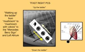
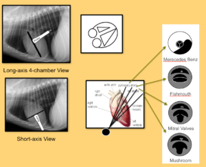
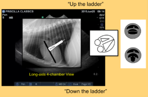
We like referring it to “walking up the ladder” and “down the ladder” of the heart, and memorizing what to expect at each level as landmarks. There is the solid muscular apex of the heart (lowest rung) and then the many pipes (circular structures) near the base of the heart (highest rung) with the mushroomed shape of the LV in between.
From Focused Ultrasound Techniques for the Small Animal Practitioner, Wiley 2014.
Once you get the LVSAV as your starting point, then you direct the probe toward the base of the heart seeing next the bright white (hyper echoic) mitral valve leaflets, followed by the so called “fishmouth view”, followed by the Mercedes Benz or “peace sign” of the LA:Ao view. By memorizing the cardiac rungs of the ladder, you get a better idea of where you are anatomically on the heart.
From Focused Ultrasound Techniques for the Small Animal Practitioner, Wiley 2014.
Once these short-axis views are repeatedly recognized and this skill is achieved, then the RV:LV on the Long-axis 4-chamber View can be attempted. At the level of the LVSAV where mitral valve leaflets or the fishmouth is viewed, the probe is rotated 90 degrees, either toward the spine or toward the sternum depending on sonographer preference. Importantly, perform the rotation the same way every time to be able to recognize where expected heart chambers should be located.
*Note, it is perfectly acceptable to do all formats, AFAST®, TFAST® and Vet BLUE® on the abdominal preset without changing screen orientation.
How is each performed?
- The LVSAV is performed at the Right Pericardial (PCS) Site by directing the probe marker toward the elbow to 4 o’clock. It is important to look at your patient’s thorax and make sure that the probe is angled through the short-axis of the heart.
- The LA:Ao Ratio is performed by imaging up the ladder fanning on the short-axis line of 4 o’clock.
- The RV:LV Ratio is found by the RVLA 4-chamber rotating the probe at the short-axis level of the mitral valves either at the windshield wipers or the fishmouth level. It is BEST to NOT look at the screen, but the probe and move it to 1 o’clock and THEN look at the screen. Yes, this really magically works so much better!
From Focused Ultrasound Techniques for the Small Animal Practitioner, Wiley 2014.
What other facets of Global FAST® are imprint markers of cardiac function?
- The importance of characterizing the Caudal Vena Cava (CVC) as it traverses the diaphragm cannot be overemphasized (FAT, flat or bounce) helping further clarify right-sided heart function and volume (your new central venous pressure is the CVC).
- The importance of using Vet BLUE® and its Dry vs. Wet Lung principles for left-sided heart function and volume cannot be over emphasized.
Video clips are available for Premium Members in our blogged FASTVet Video Library. We also have an archived 1-hour Webinar on the “TFAST® Views of the Heart” available for Premium Members. You can learn how to perform the views at our Hands On Course which is available at scheduled courses throughout the year at our Austin, Texas, FASTVet Academy or by special arrangement at your practice.
What are the Pitfalls of the TFAST® Heart Views (strongly recommend viewing the Webinar on the Ultrasound Diagnosis of Pericardial Effusion)?
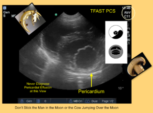
- The diagnosis of pleural and pericardial effusion should never be made along the short-axis views unless the probe is directed toward the muscular apex of the heart to look for the Bull’s Eye Sign (Focused Ultrasound Techniques for the Small Animal Practitioner, Wiley 2014). We found that non-radiologist and non-cardiologist veterinarians were sticking needles into the right ventricle (Lisciandro, JVECC 2015).
- The diagnosis of pleural and pericardial effusion should follow FASTVet tenets of using the DH View (Racetrack Sign), the TFAST Right Pericardial (Bull’s Eye Sign), and the Long-axis 4-chamber View knowing that all 4-chambers must be clearly in view and knowing the anatomical attachments of the pericardial (right atrium around apex to left atrium).
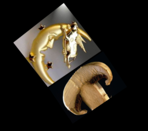
- Never diagnose at short-axis views at the level of the LV short-axis mushroom view because it is too easy to mistake the right ventricle/ right heart (man in the moon and cow jumping over the moon or the mushroom (LV) has a cap (RV) that must be appreciated) for pericardial or pleural effusion.
This information can be found in our Textbook published by Wiley 2014. To purchase the textbook, click here:
Focused Ultrasound Techniques for the Small Animal Practitioner
