The Author: Dr. Stephanie Lisciandro, DVM is a Diplomate of ACVIM and board-certified in Small Animal Internal Medicine. She has worked as a staff internist in several specialty practices, done mobile ultrasound (including complete abdominal ultrasound and echocardiography) and internal medicine specialty consulting, and telemedicine.
Overview: This case shows how Vet BLUE®, a novel, regional, pattern-based approach to lung ultrasound (and the most studied in veterinary medicine with >8 clinical studies -see References and here), helps improve/complement lung imaging over/with thoracic radiography. Take the Vet BLUE® Online Course for up-to-date information on its protocol, use for respiratory patients, including “Wet Lung” versus “Dry Lung”, its Vet BLUE® B-line scoring system, and its Vet BLUE® Visual Lung Language.
Illustrative Case: “Sasha” is a 12-year-old, spayed female, Domestic Shorthair Cat, referred for a 2-week history of inappetence and weight loss. On physical examination, she was thin with an unkempt coat and a II/VI systolic murmur. Initial evaluation included complete blood count (CBC), chemistry profile, UA, and thoracic radiographs (TXR). TXR showed a normal cardiac silhouette and a focal region of patchy infiltrates in the right caudal lung lobe. A thoracic ultrasound and Vet BLUE® lung examination were subsequently requested.


Thoracic ultrasound with Vet BLUE® revealed a focal Shred Sign (air bronchogram) in the right Vet BLUE® caudal lung region consistent with a consolidated/infiltrative lung lesion. In addition, several small (3-5 mm) nodules were also noted in the right cranial lung region. A complete detailed echocardiogram was unremarkable.

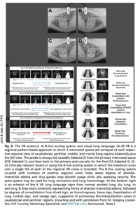
Shred Sign of Vet BLUE® in the right caudal lung region. See our Vet BLUE® Video Library for additional (cine clip) examples.
![]()
Nodule Sign of Vet BLUE® – Lung nodule (5 mm) detected with Vet BLUE® in the right cranial lung region. A Nodule Sign is defined as a hypoechoic (dark) oval or circle that has a hyperechoic (white) far border and a “pseudo B-line” (our created term, Pacholec et al. 2021) that is hyperechoic (bright white) and extends from the nodule’s far border through the far-field without fading.
Performing Vet BLUE®: Vet BLUE® follows the order of left caudodorsal lung region, to perihilar lung region, to middle lung region, and lastly the cranial lung region followed by the same regional views on the right hemithorax. A minimum of 3 intercostal spaces (ICSs) are surveyed with the highest number of B-lines over a single intercostal space found at each respective region scored as 1, 2, 3, >3, and infinity. The TFAST®-AFAST® Diaphragmatico-Hepatic (DH) View is also scored and the 9th acoustic window of Vet BLUE® providing a deep window to lung along along its pulmonary-diaphragmatic interface.
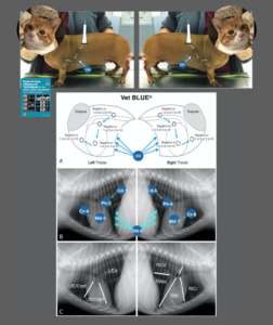
Recording Vet BLUE® Findings: A quick way to record Vet BLUE® findings is following the order of left – Cd, Ph, Md, and Cr and then right – Cd, Ph, Md, Cr always in the same order every time. The B-line score is recorded at each respective view and whether other signs of consolidation are found (Sh, Shred Sign; Ti, Tissue Sign; Nd, Nodule Sign; Wdg, Wedge Sign). We prefer left to right when we have a choice. See our goal-directed template examples in our Free Resources.
- Left Vet BLUE®: 0, 0, 0, 0. Interpretation, dry lung all views on the left side with no evidence of alveolar-interstitial lung edema or other lung surface pathology
- Right Vet BLUE®: Large Shred Sign (Sh), 0, 0, Multiple small Nodule Sign (Nd) in the right cranial lung region. Interpretation, a large region of focal consolidation (Shred Sign-air bronchogram) in the right caudal lung region. This Shred Sign measured approximately 2.7 x 1.4 cm; multiple focal nodular lesions in the right cranial lung region. *For more details on the definition of the Shred Sign and Nodule Sign, see our blogged Video Library, Proceedings, or textbook, Point-of-care Ultrasound Techniques for the Small Animal Practitioner, Wiley 2021.
Diagnostics (FNA and CT Imaging): Fine needle aspirates were performed on the right caudal lung lobe mass. Cytology was consistent with pulmonary carcinoma. Subsequently, thoracic computed tomography (CT) was performed to confirm the primary mass and evaluate for evidence of metastasis. In addition to the primary caudodorsal lung lobe mass, several small metastatic nodules were seen in the right cranial lung lobe on CT consistent with Vet BLUE® findings.
Vet BLUE® can be extremely valuable for assessing for lung pathology that is questionable or missed on TXR. Several small nodules were detected on the initial Vet BLUE® that were not evident radiographically. Therefore, the initial assessment of the thoracic ultrasound included “the nodules in the right cranial lung region are suspicious for metastasis.”
The Upshot and Clinical Implications: Moreover, in the past, the author (Stephanie Lisciandro, DVM, Dipl. ACVIM – Small Animal Internal Medicine) has previously been reluctant to perform fine needle aspirates on lung lesions that were not discrete masses on TXR along the lung periphery. However, since routinely incorporating Vet BLUE® into her thoracic imaging evaluations, fine needle aspirates are now performed far more frequently than her past 20-years of practice. “I now realize that I have another valuable method to assess lung pathology that often provides additional information to traditional radiography.”
Summary Composite of Vet BLUE® with permission from Lisciandro GR, Cageside Ultrasonography in the Emergency Room and Intensive Care Unit, Vet Clinics of North America, 2020 (Ed. Mazzafero) and modified from the textbook Point-of-care Ultrasound Techniques for the Small Animal Practitioner, Wiley 2021.
Conclusion: Vet BLUE® is an extremely valuable respiratory screening test for the evaluation of lung pathology that may be questionable or missed on TXR. There are 2 chapters devoted to the Vet BLUE® protocol, its novel regional, pattern-based approach, its Vet BLUE® B-line scoring system, and its Vet BLUE® Visual Lung Language in the 2nd edition of the textbook Point-of-care Ultrasound Techniques for the Small Animal Practitioner, Wiley 2021.
Further Resources: Our 21-hours of RACE-approved FASTVet Online Global FAST® Courses and ALL Day Global FAST® Hands-on Course (and the Webinars as part of our FASTVet Premium Membership) will provide much greater detail on how to perform, interpret, and integrate Vet BLUE® findings. Vet BLUE® is cutting-edge and a rare opportunity for those veterinarians that learn Vet BLUE® as a new core skill to be ahead of our physician colleagues.
References: Found on the FASTVet Lead page.
FAST Saves Lives! TM